

Feasibility of digitally stained multimodal confocal mosaics to simulate histopathology
- PMID: 19566342
- PMCID: PMC2929174
- DOI: 10.1117/1.3149853
Feasibility of digitally stained multimodal confocal mosaics to simulate histopathology
Abstract
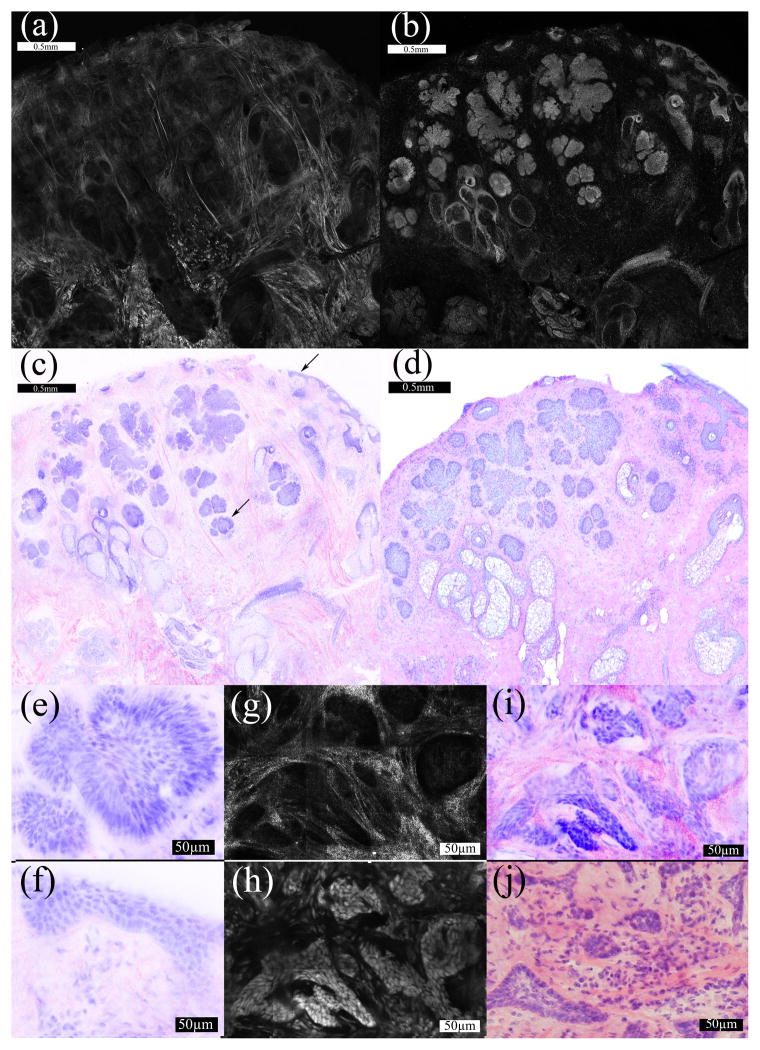
Fluorescence confocal mosaicing microscopy of tissue biopsies stained with acridine orange has been shown to accurately identify tumors and with an overall sensitivity of 96.6% and specificity of 89.2%. However, fluorescence shows only nuclear detail similar to hematoxylin in histopathology and does not show collagen or cytoplasm, which may provide necessary negative contrast information similar to eosin used in histopathology. Reflectance mode contrast is sensitive to collagen and cytoplasm without staining. To further improve sensitivity and specificity, digitally stained confocal mosaics combine confocal fluorescence and reflectance images in a multimodal pseudo-color image to mimic the appearance of histopathology with hematoxylin and eosin and facilitate the introduction of confocal microscopy into the clinical realm.
Figures
References
-
- Rajadhyaksha M, Menaker G, Flotte TJ, Dwyer PJ, Gonzalez S. Rapid confocal examination of non-melanoma cancers in skin excisions to potentially guide Mohs micrographic surgery. J Invest Dermatol. 2001;117:1137–1143. - PubMed
-
- Swanson NA. Mohs surgery: Technique, indications, applications, and the future. Arch Dermatol. 1983;119(9):761–773. - PubMed
MeSH terms
Substances
Grants and funding
LinkOut - more resources
Full Text Sources
Other Literature Sources
