Review
. 2009 Oct;10(10):1390-404.
doi: 10.1111/j.1600-0854.2009.00951.x. Epub 2009 Jun 9.

Death by committee: organellar trafficking and communication in apoptosis

Affiliations
Review

Death by committee: organellar trafficking and communication in apoptosis

Joseph E Aslan et al. Traffic. 2009 Oct.

Abstract

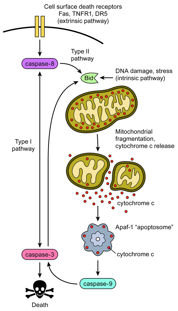
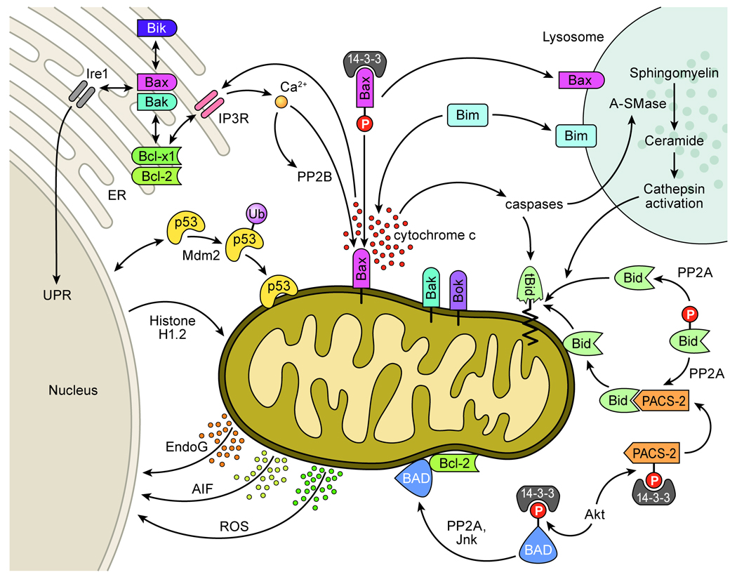
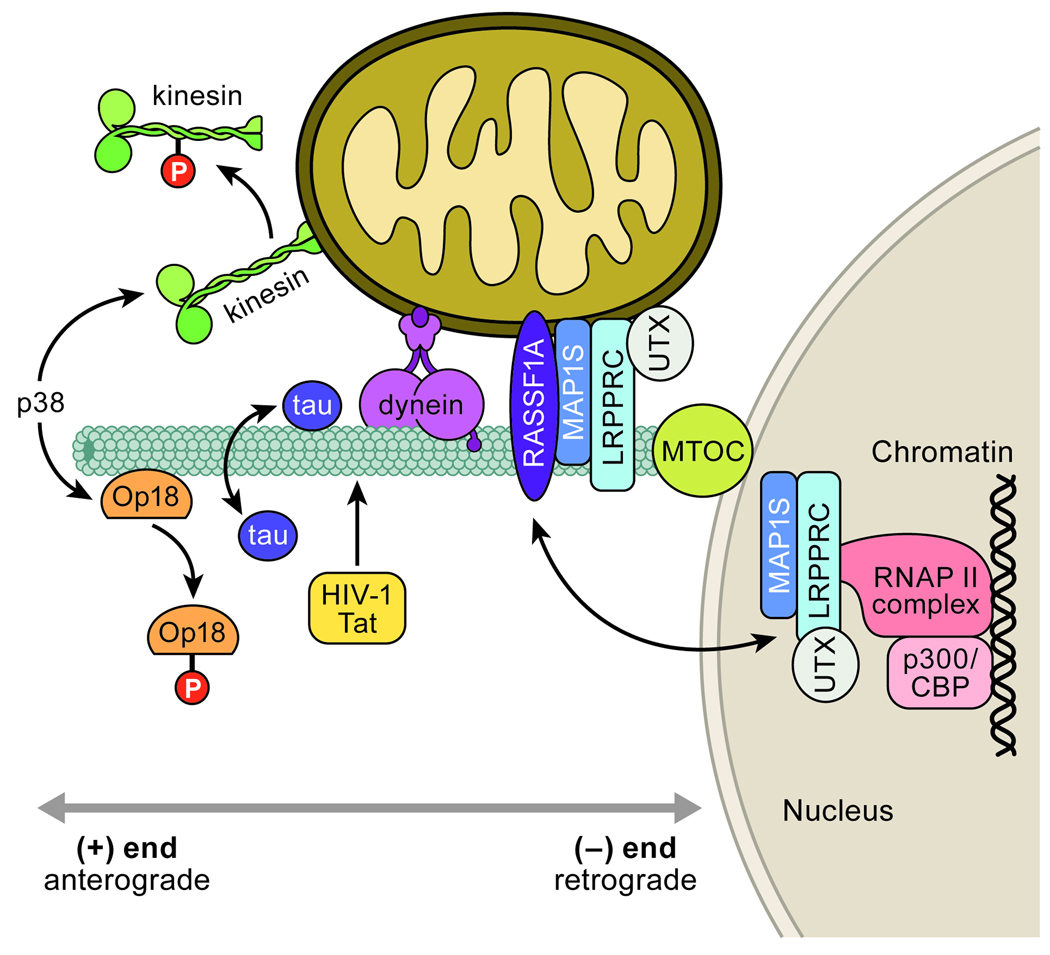
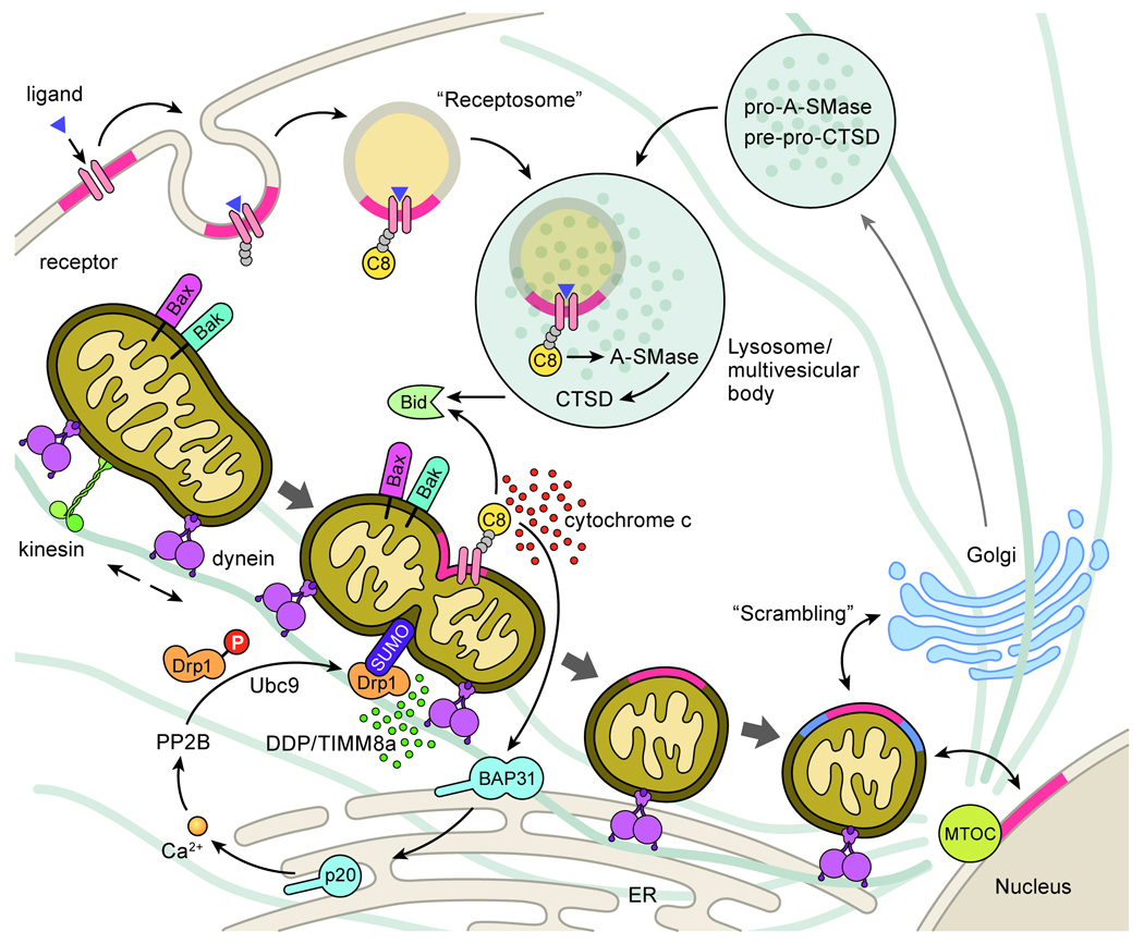
Apoptosis proceeds through a set of evolutionarily conserved processes that co-ordinate the elimination of damaged or unneeded cells. This program of cell death is carried out by organelle-directed regulators, including the Bcl-2 proteins, and ultimately executed by proteases of the caspase family. Although the biochemical mechanisms of apoptosis are increasingly understood, the underlying cell biology orchestrating programmed cell death remains enigmatic. In this review, we summarize the current understanding of Bcl-2 protein regulation and caspase activation while examining cell biological mechanisms and consequences of apoptotic induction. Organellar contributions to apoptotic induction include death receptor endocytosis, mitochondrial and lysosomal permeabilization, endoplasmic reticulum calcium release and fragmentation of the Golgi apparatus. These early apoptotic events are accompanied by stabilization of the microtubule cytoskeleton and translocation of organelles to the microtubule organizing center. Together, these phenomena establish a model of apoptotic induction whereby a cytoskeletal-dependent coalescence and 'scrambling' of organelles in the paranuclear region co-ordinates apoptotic communication, caspase activation and cell death.


Figures

Figure 1
Figure 1. Essential pathways to caspase activation and cell death
Apoptosis is initiated by internal cellular stress or extracellularly through the binding of ligands to cell surface death receptors. Type I pathways directly activate executioner caspases through initiator caspases to result in death. In Type II pathways, death signals are routed through the Bcl-2 proteins such as Bid and the mitochondria to control the release of cytochrome c. Cytosolic cytochrome c binds Apaf-1 to activate the apoptosome and caspase-9 to result in executioner caspase-3 activation and cell death.
Figure 2
Figure 2. Bcl-2 proteins modulate apoptosis at multiple organellar sites
Bcl-2 proteins converge on the mitochondria to control the release of apoptogenic factors such as cytochrome c and SMAC/DIABLO to enhance caspase activation (6). Factors such as Endonuclease G, AIF and reactive oxygen species are also released from the mitochondria to the nucleus to promote genome destruction (5, 87). Mitochondrial Bax/Bak release events are activated by Bid translocation to mitochondria and cleavage of Bid by caspases or lysosomal cathepsins (33, 43). Full-length Bid is trafficked to the mitochondria though associations with PACS-2 (59). Active cathepsins are released from lysosomes upon the translocation of Bax and Bim to lysosomes and a caspase activation o f A-SMase to produce ceramide which activates cathepsins (42, 48, 49). Apoptotic proteins such as Bad, Bax and PACS-2 are sequestered by 14-3-3 proteins and become active upon dephosphorylation and 14-3-3 release (28). Factors such as p53 and Histone H1.2 also apoptotically target the mitochondria to modulate Bax activation and apoptosis (94, 95). Bcl-2 proteins additionally regulate apoptosis via ER calcium release through IP3Rs to modulate ER-mitochondria crosstalk (73, 75, 77) and influence the UPR through interactions with Ire1 (74).
Figure 3
Figure 3. Microtubule associated motors and stabilizing proteins regulate paranuclear mitochondrial clustering and apoptosis
Apoptotic p38 phosphorylation of kinesin subunits halts anterograde traffic of mitochondria to promote dynein driven (-)-end accumulation (107). p38 phosphorylation also releases Op18 from microtubules to result in microtubule stabilization and mitochondrial clustering (116, 117). Mitochondrial aggregation and apoptosis are also influenced through microtubules by tau (83, 125) and HIV-1 Tat (126). MAP1S – LRPPRC – UXT complexes also stabilize microtubules and localize to apoptotic paranuclear mitochondria as well as the nucleus in complex with RNAPII and p300/CBP (, –124). It has been proposed that these microtubule and nucleus associated proteins coordinate both apoptotic mitochondrial clustering and chromatin remodeling events (123, 124).
Figure 4
Figure 4. Death receptor ligation promotes mitochondrial fragmentation, mitochondrial clustering, and membrane “scrambling”
Cell surface death receptors concentrate in GD3-containing lipid rafts (in red) (146). Upon death ligand engagement, receptors recruit death adaptors and are internalized with rafts to specific endosomal compartments such as “TNF receptosomes” to promote caspase-8 (C8) activation (150, 151). Receptosomes fuse with precursor hydrolase-containing Golgi-derived vesicles to form lysosomal multivesicular bodies that activate A-SMase and cathepsin D (CTSD). Caspases and cathepsins cleave Bid to promote Bax/Bak activation, cytochrome c release and caspase activation (33, 42, 43). Mitochondrial-localized caspase-8 cleaves ER localized BAP31 to a p20 fragment which promotes ER calcium release (35, 71), PP2B activation and Drp1 dephosphorylation (70) and mitochondrial fragmentation. Mitochondrial DDP/TIMM8a release (66) and Drp1 sumoylation (67, 68) promote the translocation of Drp1 to mitochondrial scission sites as mitochondria travel in the (-)-end direction (111). Bax and Bid are also recruited to these scission sites that are rich in GD3 (in red). The accumulation of fragmented mitochondria at the MTOC in proximity to the Golgi results in a caspase dependent fragmentation of the Golgi (139) and a “scrambling” of Golgi membranes (in blue) with mitochondria (50). As GD3-contatining rafts (in red) internalize to endosomes and later localize to mitochondria, it is hypothesized that mitochondria act as “cargo boats” to carry GD3 from the cell surface to the nucleus and adjacent organelles (145).

References

    1. Degterev A, Yuan J. Expansion and evolution of cell death programmes. Nat Rev Mol Cell Biol. 2008 - PubMed
    1. Eckhart L, Ballaun C, Hermann M, Vandeberg JL, Sipos W, Uthman A, Fischer H, Tschachler E. Identification of novel mammalian caspases reveals an important role of gene loss in shaping the human caspase repertoire. Mol Biol Evol. 2008 - PubMed
    1. Luthi AU, Martin SJ. The CASBAH: a searchable database of caspase substrates. Cell Death Differ. 2007;14(4):641–650. - PubMed
    1. Peter ME, Krammer PH. The CD95(APO-1/Fas) DISC and beyond. Cell Death Differ. 2003;10(1):26–35. - PubMed
    1. Ferri KF, Kroemer G. Organelle-specific initiation of cell death pathways. Nat Cell Biol. 2001;3(11):E255–E263. - PubMed

Publication types

LinkOut - more resources