. 2009 Oct;8(10):2201-11.
doi: 10.1074/mcp.M900203-MCP200. Epub 2009 Jun 29.

Proteomics characterization of cell membrane blebs in human retinal pigment epithelium cells

Affiliations

Proteomics characterization of cell membrane blebs in human retinal pigment epithelium cells

Oscar Alcazar et al. Mol Cell Proteomics. 2009 Oct.

Abstract

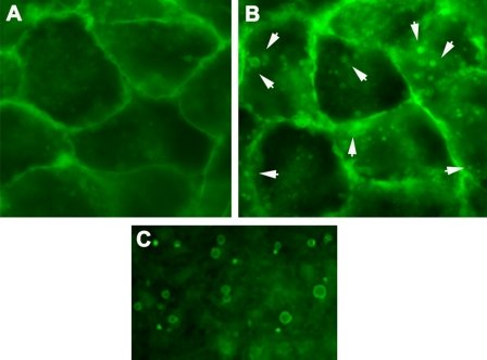
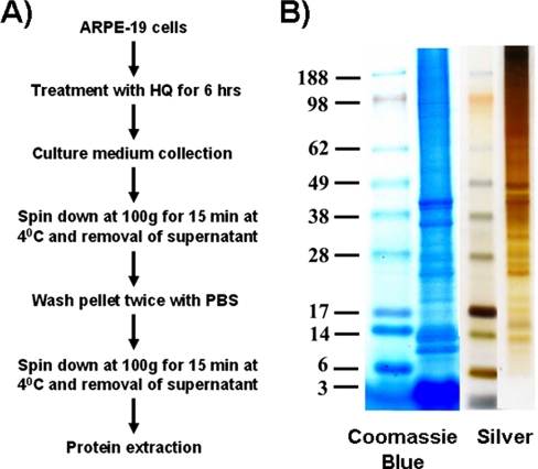
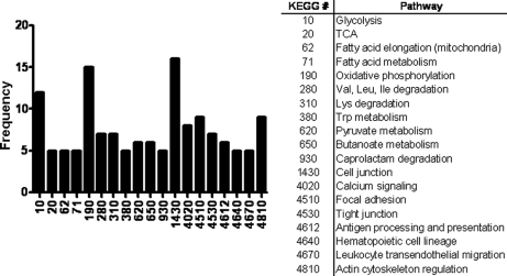
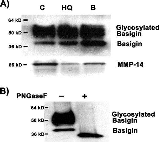
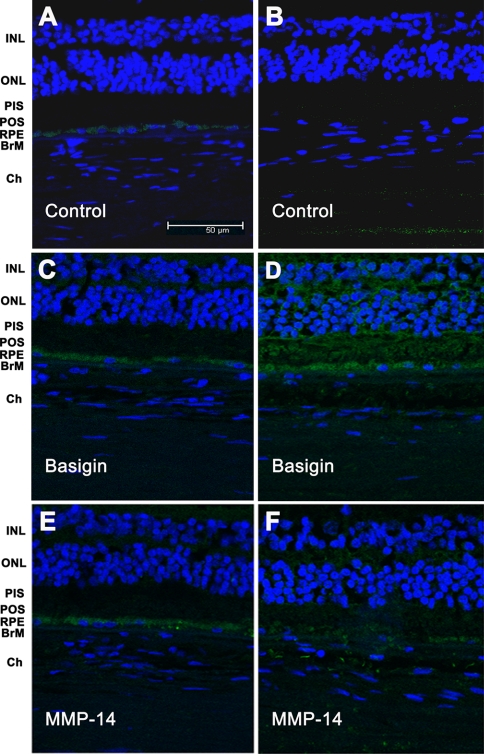
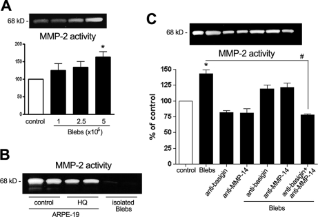
Age-related macular degeneration (AMD) is the leading cause of legal blindness among the elderly population in the industrialized world, affecting about 14 million people in the United States alone. Smoking is a major environmental risk factor for AMD, and hydroquinone is a major component in cigarette smoke. Hydroquinone induces the formation of cell membrane blebs in human retinal pigment epithelium (RPE). Blebs may accumulate and eventually contribute first to sub-RPE deposits and then drusen formation, which is a prominent histopathologic feature in eyes with AMD. As an attempt to better understand the mechanisms involved in early AMD, we sought to investigate the proteomic profile of RPE blebs. Isolated blebs were subjected to SDS-PAGE fractionation, and in-gel trypsin-digested peptides were analyzed by LC-MS/MS that lead to the identification of a total of 314 proteins. Identified proteins were predominantly involved in oxidative phosphorylation, cell junction, focal adhesion, cytoskeleton regulation, and immunogenic processes. Importantly basigin and matrix metalloproteinase-14, key proteins involved in extracellular matrix remodeling, were identified in RPE blebs and shown to be more prevalent in AMD patients. Altogether our findings suggest, for the first time, the potential involvement of RPE blebs in eye disease and shed light on the implication of cell-derived microvesicles in human pathology.


Figures

Fig. 1.
Fig. 1.
Induction of membrane blebs in ARPE-19 cells by hydroquinone-induced cellular stress. ARPE-19 cells expressing GFP at the plasma membrane (A) were exposed to HQ (100 μm) for 6 h (B). Cells were observed immediately under epifluorescence microscope (magnification, ×40). The figure shows GFP localized to the membrane and the presence of membrane blebs (white arrowheads) after HQ treatment. A detailed view of the blebs that accumulated in the conditioned medium after HQ treatment is shown (C).
Fig. 2.
Fig. 2.
Isolation of blebs. A, scheme for bleb isolation. ARPE-19 cells were treated with HQ (100 μm) for 6 h. Culture medium was collected and centrifuged at 100 × g for 15 min at 4 °C. The resulting pellet was washed twice with PBS and resuspended. The resuspended pellet was centrifuged at 100 × g for 15 min at 4 °C, and the supernatant was removed. Blebs were collected and used for protein extraction. B, representative one-dimensional gel showing the Coomassie Blue and silver stainings of resolved proteins present in ARPE-19 blebs.
Fig. 3.
Fig. 3.
Functional characterization of proteins identified in hydroquinone-induced blebs. The distribution profile of the proteins identified in hydroquinone-induced blebs is depicted according to functional categories. The KEGG database number and its corresponding metabolic pathway are shown. TCA, tricarboxylic acid cycle.
Fig. 4.
Fig. 4.
Western blot analysis of basigin and MMP-14 expression in hydroquinone-induced blebs. Protein expression was analyzed in 20 μg of total ARPE-19 cell lysate. A, basigin and MMP-14 expression in control, untreated cells (C), cells treated with 100 μm HQ for 6 h (HQ), and isolated HQ-induced membrane blebs (B). Basigin analysis shows a higher molecular mass, broad band corresponding to highly glycosylated species of basigin. B, Western blot analysis of basigin in isolated HQ-induced membrane blebs before and after digestion with the enzyme PNGaseF, which selectively removes N-linked carbohydrate residues. The blot from a representative experiment is shown. The number on the left represents the molecular mass of the protein.
Fig. 5.
Fig. 5.
Immunohistochemical analysis of basigin and MMP-14 in human retina. Retina sections from human donor eyes with no known eye disease (A, C, and E) or from human donor eyes with dry AMD (B, D, and F) were stained with either mouse polyclonal anti-basigin (C and D) or mouse monoclonal anti-MMP-14 (E and F) as indicated. Negative controls were generated by omission of the primary antibody (A and B). Secondary antibodies were coupled to Alexa Fluor 488. Nuclei were stained with 4,6-diamidino-2-phenylindole dihydrochloride. Sections were analyzed under a confocal microscope. INL, inner nuclear layer; ONL, outer nuclear layer; PIS, photoreceptor inner segments; POS, photoreceptor outer segments; Ch, choroid.
Fig. 6.
Fig. 6.
MMP-2 activity in ARPE-19 cells exposed to hydroquinone-induced blebs. Blebs were generated by incubation with 100 μm HQ for 6 h and used to treat ARPE-19 cells for 24 h. Thereafter conditioned medium was collected, and MMP-2 activity was evaluated by gelatin zymography. A, MMP-2 activity from cells (4.5 × 10⁵) incubated with increasing concentrations of blebs. B, MMP-2 activity in control, non-treated cells (C), HQ-treated cells (HQ), and isolated blebs alone (B). C, MMP-2 activity from cells incubated with blebs (5 × 10⁵ blebs/4.5 × 105 cells) in the presence or absence of anti-basigin (1:500) and anti-MMP-14 (1:500) antibodies. Top, gelatin zymogram from a representative experiment. Bottom, bar graph showing averages of results of three independent experiments. Results are expressed as mean ± S.E. #, p < 0.01; *, p < 0.05 compared with control cells. Error bars indicate standard error of the mean.

References

    1. Christen W. G., Glynn R. J., Manson J. E., Ajani U. A., Buring J. E. (1996) A prospective study of cigarette smoking and risk of age-related macular degeneration in men. JAMA 276, 1147–1151 - PubMed
    1. Clemons T. E., Milton R. C., Klein R., Seddon J. M., Ferris F. L., 3rd (2005) Risk factors for the incidence of Advanced Age-Related Macular Degeneration in the Age-Related Eye Disease Study (AREDS) AREDS report no. 19. Ophthalmology 112, 533–539 - PMC - PubMed
    1. Evans J. R. (2001) Risk factors for age-related macular degeneration. Prog. Retin. Eye Res 20, 227–253 - PubMed
    1. Khan J. C., Thurlby D. A., Shahid H., Clayton D. G., Yates J. R., Bradley M., Moore A. T., Bird A. C. (2006) Smoking and age related macular degeneration: the number of pack years of cigarette smoking is a major determinant of risk for both geographic atrophy and choroidal neovascularisation. Br. J. Ophthalmol 90, 75–80 - PMC - PubMed
    1. Vingerling J. R., Hofman A., Grobbee D. E., de Jong P. T. (1996) Age-related macular degeneration and smoking. The Rotterdam Study. Arch. Ophthalmol 114, 1193–1196 - PubMed

Publication types

MeSH terms