. 2009 Sep 15;18(18):3470-83.
doi: 10.1093/hmg/ddp291. Epub 2009 Jun 29.

Direct and indirect roles of RECQL4 in modulating base excision repair capacity

Affiliations

Direct and indirect roles of RECQL4 in modulating base excision repair capacity

Shepherd H Schurman et al. Hum Mol Genet. .

Abstract

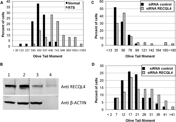
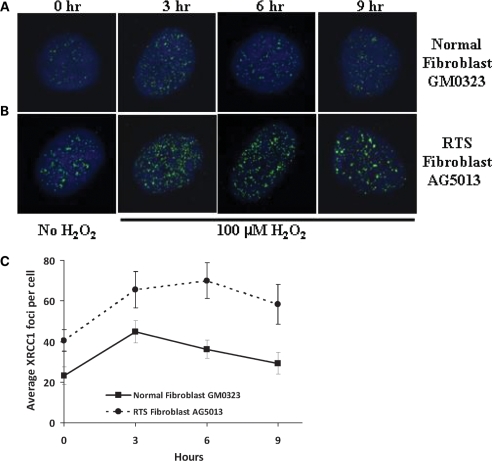
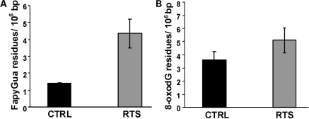
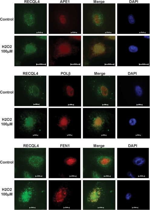
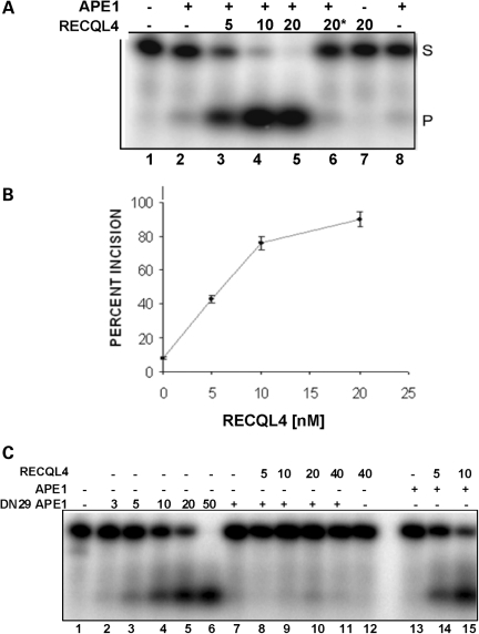
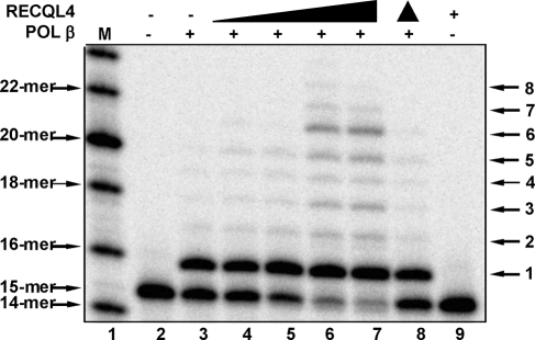
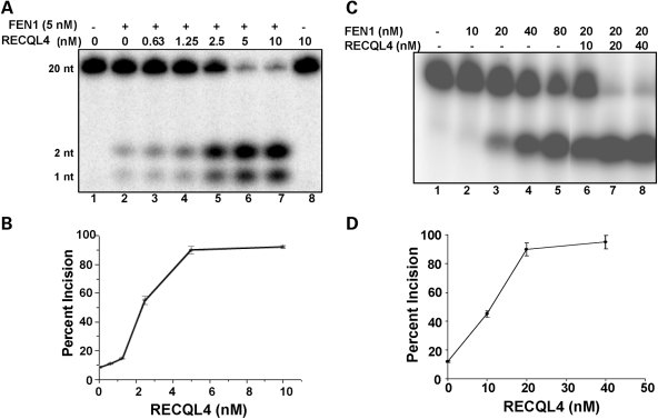
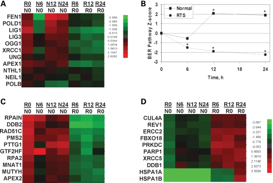
RECQL4 is a human RecQ helicase which is mutated in approximately two-thirds of individuals with Rothmund-Thomson syndrome (RTS), a disease characterized at the cellular level by chromosomal instability. BLM and WRN are also human RecQ helicases, which are mutated in Bloom and Werner's syndrome, respectively, and associated with chromosomal instability as well as premature aging. Here we show that primary RTS and RECQL4 siRNA knockdown human fibroblasts accumulate more H(2)O(2)-induced DNA strand breaks than control cells, suggesting that RECQL4 may stimulate repair of H(2)O(2)-induced DNA damage. RTS primary fibroblasts also accumulate more XRCC1 foci than control cells in response to endogenous or induced oxidative stress and have a high basal level of endogenous formamidopyrimidines. In cells treated with H(2)O(2), RECQL4 co-localizes with APE1, and FEN1, key participants in base excision repair. Biochemical experiments indicate that RECQL4 specifically stimulates the apurinic endonuclease activity of APE1, the DNA strand displacement activity of DNA polymerase beta, and incision of a 1- or 10-nucleotide flap DNA substrate by Flap Endonuclease I. Additionally, RTS cells display an upregulation of BER pathway genes and fail to respond like normal cells to oxidative stress. The data herein support a model in which RECQL4 regulates both directly and indirectly base excision repair capacity.


Figures

Figure 1.
Figure 1.
Comet assay of RTS and RECQL4 knockdown cells treated with H2O2. (A) Comet assay for RTS fibroblasts. Exponentially growing normal, MRC5, or RTS, AG5013, cells were treated with 500 µM H2O2 for 15 min on ice, and comet assay was performed as described in Materials and Methods. (B) Western blot analysis of U2OS cells treated with RECQL4 siRNA. Blot was probed with the indicated antibody. Western blot of U2OS whole cell extracts showing the knockdown effects of RECQL4 siRNA: (1) No treatment; (2) Negative control siRNA; (3) RECQL4 siRNA; (4) Purified RECQL4 protein (∼5 ng). (C) Comet assay for RECQL4 knockdown U2OS cells treated with H2O2. RECQL4 knockdown U2OS cells were treated with 500 µM H2O2 for 15 min and comet assay was performed as described in Materials and Methods. (D) Comet assay for RECQL4 knockdown cells with 3 h recovery after treatment with H2O2. Same as in (C), except cells were incubated for 3 h in fresh medium lacking H2O2 before comet assay was performed. Comet assay data was analyzed using Komet 5.5 software. Shown is the percentage of cells with the observed Olive Tail moment values. In order to graphically represent the OTMs, the data were binned into 10–12 segments and, unless otherwise noted, the median of the bin is displayed on the X-axis.
Figure 2.
Figure 2.
XRCC1 foci in RTS cells treated with H2O2. The GM00323 normal (A) and AG5013 RTS fibroblast (B) cell lines were incubated in the absence (0 h) or presence of 100 µM H2O2 for 15 min, and then fixed 3, 6 or 9 h after H2O2 treatment. Cells were stained with rabbit anti-XRCC1 antibody as described in Materials and Methods. Images were captured with a Nikon TE2000 microscope at 600× magnification with five laser imaging modules and a CCD camera (Hamamatsu). (C) Average number of foci observed per cell, minimum number of cells analyzed for each time point was 10 cells. Significant P-values between the normal and RTS cells were 0.024, 0.044, 0.0031 and 0.001 for the 0, 3, 6 and 9 h points, respectively.
Figure 3.
Figure 3.
Endogenous levels of FapyG and 8-oxoG in RTS cells. DNA was isolated from normal control, GM00323 and RTS fibroblasts, AG5013, digested with P1 nuclease and alkaline phosphatase, then analyzed by GC-MS for FapyG (A) or 8-oxoG (B) as described in Materials and Methods. Values indicated are modified residues per 106 bases.
Figure 4.
Figure 4.
Immunohistochemical localization of RECQL4, APE1, POL β and FEN1 in HeLa cells treated with H2O2. HeLa cells were incubated in the presence or absence of 100 µM H2O2 for 30 min and then fixed. Cells were stained for DAPI, RECQL4 and APE1, POL β or FEN1, as described in Materials and Methods.
Figure 5.
Figure 5.
Effect of RECQL4 on APE1 endonuclease activity. (A) APE1 incision assays were carried out using a 34 bp dsDNA substrate containing a tetrahydrofuran at position 16. APE1 (20 pg; ∼55 fM) was added as indicated before addition of the indicated amount of RECQL4. Asterisk indicates heat inactivation. (B) APE incision assays were quantified and average percent incision was calculated using data from three experiments. (C) AP incision assays were carried out with DN29 APE1 in the presence or absence of RECQL4. Plus symbol indicates addition of 2.5 pg DN29 APE1 or 20 pg of APE1, prior to addition of the indicated amount of RECQL4.
Figure 6.
Figure 6.
Effect of RECQL4 on POL β DNA synthesis activity. Increasing amounts of RECQL4 protein were added to reaction mixtures containing 12.5 nM DNA and 1 nM POL β where indicated. RECQL4 concentrations were 0, 0.75, 1.5, 3 and 6 nM. Filled triangle indicates heat inactivation. Assays were performed as described in Materials and Methods.
Figure 7.
Figure 7.
Effect of RECQL4 on FEN1 flap incision activity. FEN1 incision activity was measured using a 1-nucleotide (A and B) or 10-nucleotide (C and D) flap DNA substrate as described in Materials and Methods. The indicated amount of RECQL4 was added. (B and D) FEN1 incision assays were quantified and mean ± standard error was calculated using data from at least two experiments.
Figure 8.
Figure 8.
BER pathway analysis of microarray data. (A) Clustering of RNA expression patterns for select BER genes observed after no treatment (R0/N0) or after treatment with 25 µM menadione and harvested at 6, 12 or 24 h later. (B) Relative BER pathway gene expression changes for 11 BER proteins, OGG1, NTH1, NEIL1, UNG, APEX1, POLB, POLD, FEN1, LIG1, LIG3 and XRCC1, were analyzed and are displayed. The relative RTS versus normal signal is plotted as a function of the time when the samples were harvested. All points were statistically significant (*) except the 6 h point in the normal cells. A P-value of 0.05 was considered statistically significant. (C and D) Cluster analysis of the top 10 most up- or down-regulated DNA repair genes after menadione treatment. R0/N0, ratio of z-scores of RTS to normal cells. Nx/N0 or Rx/N0, ratio of z-scores of normal (N) or RTS (R) relative to z-score at time zero (N0 or R0), where x represents the time of harvest after menadione treatment, 6, 12 or 24 h.

References

    1. Wang L.L., Levy M.L., Lewis R.A., Chintagumpala M.M., Lev D., Rogers M., Plon S.E. Clinical manifestations in a cohort of 41 Rothmund-Thomson syndrome patients. Am. J. Med. Genet. 2001;102:11–17. - PubMed
    1. Ying K.L., Oizumi J., Curry C.J. Rothmund-Thomson syndrome associated with trisomy 8 mosaicism. J. Med. Genet. 1990;27:258–260. - PMC - PubMed
    1. Wang L.L., Gannavarapu A., Kozinetz C.A., Levy M.L., Lewis R.A., Chintagumpala M.M., Ruiz-Maldanado R., Contreras-Ruiz J., Cunniff C., Erickson R.P., et al. Association between osteosarcoma and deleterious mutations in the RECQL4 gene in Rothmund-Thomson syndrome. J. Natl Cancer Inst. 2003;95:669–674. - PubMed
    1. Siitonen H.A., Kopra O., Kaariainen H., Haravuori H., Winter R.M., Saamanen A.M., Peltonen L., Kestila M. Molecular defect of RAPADILINO syndrome expands the phenotype spectrum of RECQL diseases. Hum. Mol. Genet. 2003;12:2837–2844. - PubMed
    1. Van Maldergem L., Siitonen H.A., Jalkh N., Chouery E., De Roy M., Delague V., Muenke M., Jabs E.W., Cai J., Wang L.L., et al. Revisiting the craniosynostosis-radial ray hypoplasia association: Baller-Gerold syndrome caused by mutations in the RECQL4 gene. J. Med. Genet. 2006;43:148–152. - PMC - PubMed

Publication types

MeSH terms