. 2009 Jun;21(6):1769-80.
doi: 10.1105/tpc.109.067785. Epub 2009 Jun 30.

The PLASTID DIVISION1 and 2 components of the chloroplast division machinery determine the rate of chloroplast division in land plant cell differentiation

Affiliations

The PLASTID DIVISION1 and 2 components of the chloroplast division machinery determine the rate of chloroplast division in land plant cell differentiation

Kumiko Okazaki et al. Plant Cell. 2009 Jun.

Abstract

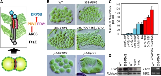
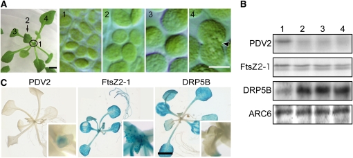
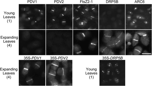
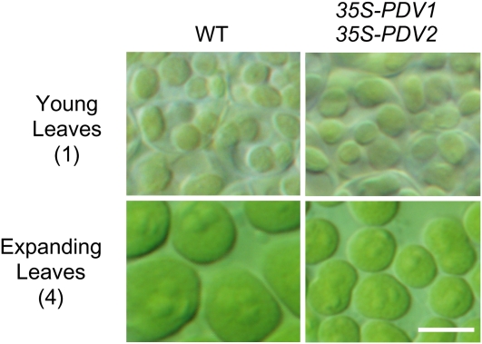
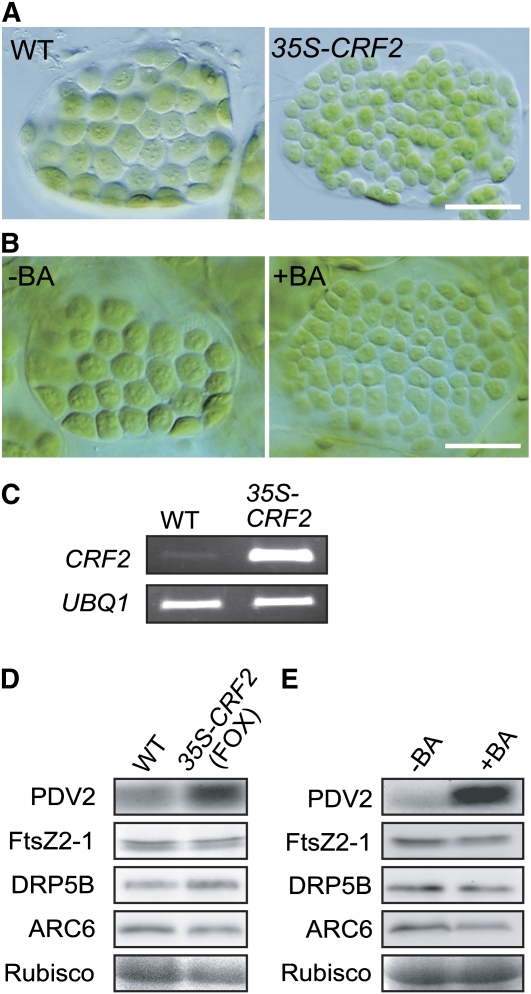
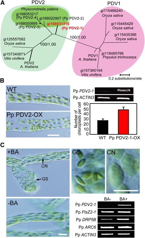
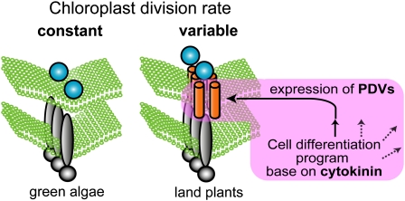
In most algae, the chloroplast division rate is held constant to maintain the proper number of chloroplasts per cell. By contrast, land plants evolved cell and chloroplast differentiation systems in which the size and number of chloroplasts change along with their respective cellular function by regulation of the division rate. Here, we show that PLASTID DIVISION (PDV) proteins, land plant-specific components of the division apparatus, determine the rate of chloroplast division. Overexpression of PDV proteins in the angiosperm Arabidopsis thaliana and the moss Physcomitrella patens increased the number but decreased the size of chloroplasts; reduction of PDV levels resulted in the opposite effect. The level of PDV proteins, but not other division components, decreased during leaf development, during which the chloroplast division rate also decreased. Exogenous cytokinins or overexpression of the cytokinin-responsive transcription factor CYTOKININ RESPONSE FACTOR2 increased the chloroplast division rate, where PDV proteins, but not other components of the division apparatus, were upregulated. These results suggest that the integration of PDV proteins into the division machinery enabled land plant cells to change chloroplast size and number in accord with the fate of cell differentiation.


Figures

Figure 1.
Figure 1.
Overexpression of PDV1 and PDV2 Increases the Number and Decreases the Size of Chloroplasts. (A) Diagram showing the pathway of chloroplast division complex assembly (Yang et al., 2008). Only the known division site–localized components are shown. FtsZ, homolog of the tubulin-like bacterial division GTPase, self-assembles into a ring structure at the stromal side of the division site (Vitha et al., 2001; Kuroiwa et al., 2002). The positioning of the FtsZ ring mid-chloroplast is regulated by MinD, MinE, ARC3, and MCD1 (Colletti et al., 2000; Itoh et al., 2001; Shimada et al., 2004; Maple et al., 2007; Nakanishi et al., 2009). FtsZ filaments are stabilized by cyanobacteria-descended inner envelope spanning protein ARC6 (Vitha et al., 2003) through interaction with FtsZ (Glynn et al., 2008). ARC6 recruits the outer envelope spanning proteins PDV1 and PDV2 through direct interaction with PDV2 (Glynn et al., 2008). PDV1 and PDV2 are required for recruitment of the cytosolic dynamin-related GTPase DRP5B at the division site (Miyagishima et al., 2006). FtsZ and ARC6 are descendants of cyanobacterial cell division machinery, while DRP5B is derived from eukaryotic membrane fission machinery, and PDV proteins are specific to land plants. (B) Chloroplasts of the wild type, transgenic plants overexpressing PDV1, PDV2, and both PDV1 and PDV2 (35S-PDV1, 35S-PDV2, and 35S-PDV1 35S-PDV2) , and pdv2/PDV2 and pdv2/pdv2 T-DNA insertional mutants. Tips of the first true leaves were cut from ∼3-week-old plants grown on agar plates. Single leaf mesophyll cells observed by Nomarski optics are shown. There are no visible differences in growth among these lines. Bar = 20 μm. (C) Statistical comparison of the number of chloroplasts per mesophyll cell. Error bars represent sd (n = 50 cells). (D) Immunoblot and RT-PCR analyses showing the levels of PDV2 protein and the PDV1 transcript. The same amount of total protein extracted from rosettes was analyzed by anti-PDV2 antibodies. The ribulose-1,5-bisphosphate carboxylase/oxygenase (Rubisco) large subunit stained by Ponceau S is shown as the loading control. Three biological replicates showed the same result for the immunoblot analyses. Total RNA extracted from rosettes was used for RT-PCR to examine the PDV1 transcript level. UBQ1 was used as the internal control. The number of PCR cycles was 28 for PDV1 and 26 for UBQ1. The signals were estimated by ethidium bromide staining. Two biological replicates showed the same result. [See online article for color version of this figure.]
Figure 2.
Figure 2.
The PDV2 Level, but Not the Other Chloroplast Division Protein Levels, Decreases in Accord with the Increase of Chloroplast Size during Leaf Development. (A) Change of chloroplast size during leaf development. Chloroplasts in a young emerging leaf (1) and expanding leaves (2 to 4) of the wild-type plant were observed by Nomarski optics. Chloroplasts are observably still dividing in the first true leaf (4, indicated by arrow). Bars = 2 mm (left) and 5 μm (right, panels 1 to 4). (B) Immunoblot analyses showing the levels of chloroplast division proteins during leaf development. PDV2 and FtsZ2-1 are detected by anti-PDV2 and anti-FtsZ2-1 antibodies, respectively. DRP5B and ARC6 levels were analyzed by anti-GFP antibodies using wild-type plants expressing GFP-DRP5B and ARC6-GFP by their respective promoters. The same amount of protein extracted from leaves corresponding to stages 1 to 4 (indicated in [A], a sample of stage 1 including both the young emerging leaves and shoot apexes) was loaded in each lane. Three biological replicates showed the same result. (C) Histochemical GUS staining of PDV2 promoter-GUS, FtsZ2-1 promoter-GUS, and DRP5B promoter-GUS transgenic plants. Approximately three-week-old plants grown on agar plates were stained. Magnified images of the centers of the rosettes are also shown (insets). ARC6 promoter-GUS transgenic plants were also prepared, but we could not obtain a staining signal. Three independent transgenic lines for each promoter showed the same results. Bar = 3 mm.
Figure 3.
Figure 3.
Division Site Localization of PDV1, PDV2, FtsZ, DRP5B, and ARC6 during Leaf Development. Fluorescence microscopy showing the localization of chloroplast division proteins during leaf development. GFP-PDV1, GFP-PDV2, FtsZ-GFP, GFP-DRP5B, and ARC6-GFP expressed by their respective promoters were observed in emerging (top panels) and expanding (middle panels) leaves corresponding to stages 1 and 4 in Figure 2. GFP-PDV1, GFP-PDV2, and GFP-DRP5B were overexpressed in wild-type plants, and the localization was observed by fluorescence microscopy (bottom panels). In emerging (35S-DRP5B) and expanding (35S-PDV1 and 35S-PDV2) leaves corresponding to stages 1 and 4 in Figure 2, these proteins localize at the chloroplast division site. Three independent transgenic lines for each GFP fusion showed the same results. Bars = 5 μm.
Figure 4.
Figure 4.
Effect of PDV1 and PDV2 Overexpression on the Young Emerging Leaves and Expanding Leaves. Chloroplasts of the wild type and PDV1 and PDV2 overexpressers in emerging and expanding leaves corresponding to stages 1 and 4 in Figure 2 A. Bar = 5 μm. [See online article for color version of this figure.]
Figure 5.
Figure 5.
CRF2 Overexpression or Cytokinin Treatment Increases the PDV2 Level and Increases the Number and Decreases the Size of Chloroplasts. (A) Chloroplasts of wild-type and transgenic plants overexpressing CRF2 (35S-CRF2) in single leaf mesophyll cells. Tips of the first true leaves were cut from ∼3-week-old plants grown on agar plates. Bar = 20 μm. (B) Effect of cytokinin treatment on the size and number of chloroplasts. Chloroplasts in single mesophyll cells of the cotyledon are shown. Wild-type seeds were germinated and grown for 10 d on agar plates with (+BA) or without (−BA) 5 μM BA. Bar = 20 μm. (C) RT-PCR analyses showing the CRF2 transcript was increased in 35S-CRF2 transgenic plants. Total RNA extracted from rosettes was used for RT-PCR. UBQ1 was used as the internal control. The number of PCR cycles was 28 for CRF2 and 26 for UBQ1. The signals were estimated by ethidium bromide staining. Two biological replicates showed the same result. (D) Immunoblot analyses comparing the levels of the chloroplast division proteins between the wild type and CRF2 overexpresser. The same amount of total protein extracted from ∼3-week-old russets was loaded in each lane. Rubisco large subunit stained by Ponceau S is shown as the loading control. Three biological replicates showed the same result. (E) Immunoblot analyses comparing the levels of the chloroplast division proteins between wild-type seedlings germinated on medium with (+BA) or without (−BA) BA. The same amount of total protein extracted from 10-d-old seedlings was loaded in each lane. PDV2, FtsZ2-1, GFP-DRP5B, and ARC6-GFP were detected as in Figure 2B. Three biological replicates showed the same result. [See online article for color version of this figure.]
Figure 6.
Figure 6.
PDV2 also Rate-Determines Chloroplast Division in the Moss P. patens. (A) Phylogenetic relationships in the PDV family of proteins. The amino acid sequences were collected from the National Center for Biotechnology Information database of nonredundant protein sequences. The GI numbers of the respective amino acid sequences are indicated. The tree shown is the maximum likelihood tree constructed by the RaxML program (Stamatakis, 2006) based on the alignment of 144 amino acid residues of 13 proteins. The numbers at the selected nodes are local bootstrap values (left) and Bayesian posterior probabilities (right) calculated by the maximum-likelihood method and Bayesian inference analyses, respectively. (B) Number of chloroplasts per chloronema cells of the wild type and Pp PDV2-1 overexpresser (Pp PDV2-OX) (n = 30 cells). In P. patens, protonema cells are classified into chloronema and caulonema. Chloronema, which contains round chloroplasts, develops to caulonema, which contains spindle-shaped chloroplasts. An increase in number of chloroplasts was also observed in caulonemal cells of the Pp PDV2-1 overexpresser. RT-PCR analyses showing the Pp PDV2-1 transcript is increased in the transgenic line. Total RNA extracted from protonemal colonies was used for RT-PCR. Pp ACTIN3 was used as the internal control. The number of PCR cycles was 28 for Pp PDV2-1 and 28 for Pp ACTIN3. The signals were estimated by ethidium bromide staining. The same results were obtained in four independent transformants. Bar = 10 μm. (C) Effect of cytokinin treatment on chloroplasts and expression of chloroplast division genes. RT-PCR analyses comparing transcript levels of chloroplast division genes between cells grown on medium with (+BA) or without (−BA) BA. Four-day-old protonemal cells were transferred onto medium with or without 5 μM BA and grown for 4 d. Pp ACTIN3 was used as the internal control. The number of PCR cycles was 28 for each gene. The signals were estimated by ethidium bromide staining. Two biological replicates showed the same result. CN, caulonema; GS, gametophore shoot apical cell of a bud induced by cytokinin. Bars = 10 μm. [See online article for color version of this figure.]
Figure 7.
Figure 7.
Schematic Representation of PDV-Mediated Control of the Chloroplast Division Rate That Evolved in Land Plants. In a common ancestor of land plants, PDV proteins became inserted into the chloroplast division apparatus descended from green algae. PDV mediate recruitment of the eukaryotic DRP5B to the division site in which cyanobacteria-descended FtsZ and ARC6 have been assembled. Levels of PDV expression are regulated by a cytokinin-dependent cell differentiation program, and the PDV level determines the rate of division site constriction. [See online article for color version of this figure.]

Comment in

References

    1. Altschul, S.F., Madden, T.L., Schaffer, A.A., Zhang, J., Zhang, Z., Miller, W., and Lipman, D.J. (1997). Gapped BLAST and PSI-BLAST: A new generation of protein database search programs. Nucleic Acids Res. 25 3389–3402. - PMC - PubMed
    1. Boffey, S.A., and Lloyd, D. (1988). Division and Segregation of Organelles. (Cambridge, UK: Cambridge University Press).
    1. Clough, S.J., and Bent, A.F. (1998). Floral dip: A simplified method for Agrobacterium-mediated transformation of Arabidopsis thaliana. Plant J. 16 735–743. - PubMed
    1. Colletti, K.S., Tattersall, E.A., Pyke, K.A., Froelich, J.E., Stokes, K.D., and Osteryoung, K.W. (2000). A homologue of the bacterial cell division site-determining factor MinD mediates placement of the chloroplast division apparatus. Curr. Biol. 10 507–516. - PubMed
    1. Gao, H., Kadirjan-Kalbach, D., Froehlich, J.E., and Osteryoung, K.W. (2003). ARC5, a cytosolic dynamin-like protein from plants, is part of the chloroplast division machinery. Proc. Natl. Acad. Sci. USA 100 4328–4333. - PMC - PubMed

Publication types

MeSH terms

Associated data

LinkOut - more resources