. 2009 Jun;21(6):1830-45.
doi: 10.1105/tpc.109.066670. Epub 2009 Jun 30.

The multifunctional enzyme CYP71B15 (PHYTOALEXIN DEFICIENT3) converts cysteine-indole-3-acetonitrile to camalexin in the indole-3-acetonitrile metabolic network of Arabidopsis thaliana

Affiliations

The multifunctional enzyme CYP71B15 (PHYTOALEXIN DEFICIENT3) converts cysteine-indole-3-acetonitrile to camalexin in the indole-3-acetonitrile metabolic network of Arabidopsis thaliana

Christoph Böttcher et al. Plant Cell. 2009 Jun.

Abstract

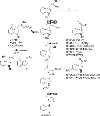
Accumulation of camalexin, the characteristic phytoalexin of Arabidopsis thaliana, is induced by a great variety of plant pathogens. It is derived from Trp, which is converted to indole-3-acetonitrile (IAN) by successive action of the cytochrome P450 enzymes CYP79B2/B3 and CYP71A13. Extracts from wild-type plants and camalexin biosynthetic mutants, treated with silver nitrate or inoculated with Phytophthora infestans, were comprehensively analyzed by ultra-performance liquid chromatography electrospray ionization quadrupole time-of-flight mass spectrometry. This metabolomics approach was combined with precursor feeding experiments to characterize the IAN metabolic network and to identify novel biosynthetic intermediates and metabolites of camalexin. Indole-3-carbaldehyde and indole-3-carboxylic acid derivatives were shown to originate from IAN. IAN conjugates with glutathione, gamma-glutamylcysteine, and cysteine [Cys(IAN)] accumulated in challenged phytoalexin deficient3 (pad3) mutants. Cys(IAN) rescued the camalexin-deficient phenotype of cyp79b2 cyp79b3 and was itself converted to dihydrocamalexic acid (DHCA), the known substrate of CYP71B15 (PAD3), by microsomes isolated from silver nitrate-treated Arabidopsis leaves. Surprisingly, yeast-expressed CYP71B15 also catalyzed thiazoline ring closure, DHCA formation, and cyanide release with Cys(IAN) as substrate. In conclusion, in the camalexin biosynthetic pathway, IAN is derivatized to the intermediate Cys(IAN), which serves as substrate of the multifunctional cytochrome P450 enzyme CYP71B15.


Figures

Figure 1.
Figure 1.
Biosynthesis of Camalexin and Related Indolic Compounds; Published Data Are Summarized (Glawischnig, 2007; Nafisi et al., 2007). Indole-3-carbaldehyde (I3CHO) and indole-3-carboxylic acid (I3CO2H) derivatives have been identified previously (Hagemeier et al., 2001; Tan et al., 2004; Bednarek et al., 2005), but their biosynthetic origin was unclear. I3CHO was also suggested as biosynthetic intermediate in camalexin biosynthesis (Zook and Hammerschmidt, 1997). ESP, epithio-specifier protein.
Figure 2.
Figure 2.
Extraction of Features Originating from Metabolites Up- and Downstream of PAD3 by Comparative Nontargeted Analyses of LC/Positive Ion ESI-QTOF-MS Metabolite Profiles of Wild Types and Biosynthetic Mutants. Venn diagrams in each row are based on alignments of 48 LC/MS analyses each [biological replicate (2) × technical replicate (4) × treatment (2) × genotype (3)]. Specific feature sets used for construction of Venn diagrams are derived from pairwise comparisons shown adjacent to each circle. A plus sign indicates a treated genotype and a minus sign a mock-treated one. A feature is assumed as differential in a pairwise comparison if it is detectable in 75% of the technical replicates and it displays at least a twofold change at a significance level of P < 0.05 in all biological replicates. After spraying silver nitrate, 62 features were mapped to putative leaf metabolites downstream of PAD3 ([A], left) and 126 upstream of PAD3 ([A], right). After inoculation with P. infestans, 88 (212) features were assigned to putative leaf metabolites downstream (upstream) of PAD3 (B). Nontargeted metabolite profiling of spore suspension droplets recollected from leaves revealed 65 (57) features associated to secreted metabolites downstream (upstream) PAD3 (C).
Figure 3.
Figure 3.
Structures of Identified and Putatively Annotated Metabolites Detected Down- and Upstream of PAD3. For annotation level of each metabolite, see Tables 1 and 2. Note that in case of compounds 2 to 4, 20, and 21, the position of the substituent on the indole ring could not be determined by mass spectrometry. Analogously, the position of the hexose moiety (Hex) in 19 is undefined. For the complete mass spectrometric characterization and additional remarks on identification, see Supplemental Data Set 2 online.
Figure 4.
Figure 4.
Relative quantification of annotated metabolites within a single independent experiment using UPLC/ESI(+)-QTOF-MS and quantifier ions from Tables 1 and 2. Levels of compounds 1-28 in leaf tissue after abiotic (silver nitrate) and biotic (P. infestans) stress application are shown in (A) and (B), respectively. Levels of compounds detected in recollected spore suspension droplets are shown in (C). Relative quantification in positive and negative ion mode for three independent experiments can be found in Supplemental Data Set 3.
Figure 5.
Figure 5.
In Vivo Feeding of Metabolic Intermediates to Arabidopsis Rosette Leaves. Camalexin concentrations of individual leaf samples are shown on a logarithmic scale. Metabolic complementation of cyp79b2 cyp79b3 by 0.25 mM Cys(IAN) was observed. Pretreatment of leaves with silver nitrate resulted in ∼16-fold increase in camalexin concentration in comparison to untreated leaves. In cyp79b2 cyp79b3, no camalexin was detected without Cys(IAN) addition. Small but highly significant (analysis of variance, P < 0.001) Cys(IAN)-dependent camalexin formation was observed in silver nitrate–treated pad3 mutants. The bars represent the mean value for each treatment. lod, limit of detection.
Figure 6.
Figure 6.
In Vivo Feeding of Metabolic Intermediates to Nontreated Rosette Leaves of Col-0 or 35S:CYP71B15 (35S:PAD3). The compound fed is shown on the abscissa. Camalexin concentrations of individual leaf samples are shown. The bars represent the mean value for each treatment.
Figure 7.
Figure 7.
Kinetic Properties of CYP71B15 Expressed in the Yeast Strain WAT11 with Cys(IAN) as Substrate. Turnover rates of Cys(IAN) with (square, solid line) and without (triangles, dashed line) NADPH as cosubstrate were calculated, based on the synthesis of DHCA and camalexin (A) or of cyanide (B).
Figure 8.
Figure 8.
Elucidation of the IAN-dependent network by combination of in vivo feeding experiments and LC/MS-based metabolite profiling. (A) Metabolization of 6-fluoro-IAN after feeding to silver nitrate–challenged pad3 leaves. (B) Relative quantification of IAN-derived metabolites after feeding of water, IAN, or 6F-IAN to nontreated or silver nitrate–treated pad3 leaves. For quantifier ions, retention times, and absolute peak area, refer to Supplemental Table 3 online.
Figure 9.
Figure 9.
Model of the IAN-Dependent Indole-3-Carboxylate/Camalexin Metabolic Network. Solid and dashed arrows indicate known and putative metabolic transformations, respectively. Transformations marked by outlined arrows were predominantly observed in the pad3 mutant background.

References

    1. Bednarek, P., Pislewska-Bednarek, M., Svatos, A., Schneider, B., Doubsky, J., Mansurova, M., Humphry, M., Consonni, C., Panstruga, R., Sanchez-Vallet, A., Molina, A., and Schulze-Lefert, P. (2009). A glucosinolate metabolism pathway in living plant cells mediates broad-spectrum antifungal defense. Science 323 101–106. - PubMed
    1. Bednarek, P., Schneider, B., Svatos, A., Oldham, N.J., and Hahlbrock, K. (2005). Structural complexity, differential response to infection, and tissue specificity of indolic and phenylpropanoid secondary metabolism in Arabidopsis roots. Plant Physiol. 138 1058–1070. - PMC - PubMed
    1. Blum, R., Beck, A., Korte, A., Stengel, A., Letzel, T., Lendzian, K., and Grill, E. (2007). Function of phytochelatin synthase in catabolism of glutathione-conjugates. Plant J. 49 740–749. - PubMed
    1. Böttcher, C., von Roepenack-Lahaye, E., Schmidt, J., Schmotz, C., Neumann, S., Scheel, D., and Clemens, S. (2008). Metabolome analysis of biosynthetic mutants reveals a diversity of metabolic changes and allows identification of a large number of new compounds in Arabidopsis. Plant Physiol. 147 2107–2120. - PMC - PubMed
    1. Boyes, D.C., Zayed, A.M., Ascenzi, R., McCaskill, A.J., Hoffman, N.E., Davis, K.R., and Gorlach, J. (2001). Growth stage-based phenotypic analysis of Arabidopsis: A model for high throughput functional genomics in plants. Plant Cell 13 1499–1510. - PMC - PubMed

Publication types

MeSH terms