

Rational engineering of type II restriction endonuclease DNA binding and cleavage specificity
- PMID: 19567736
- PMCID: PMC2731914
- DOI: 10.1093/nar/gkp535
Rational engineering of type II restriction endonuclease DNA binding and cleavage specificity
Abstract
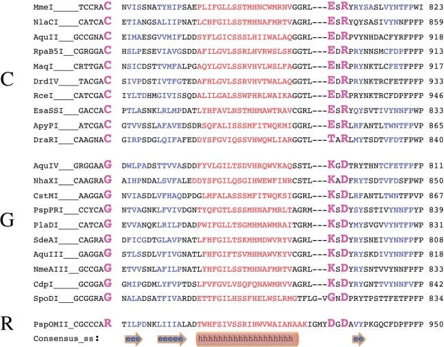
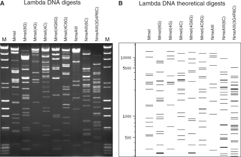
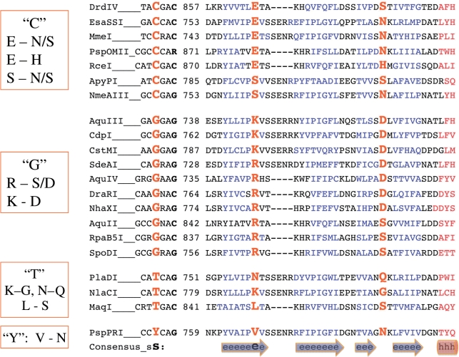
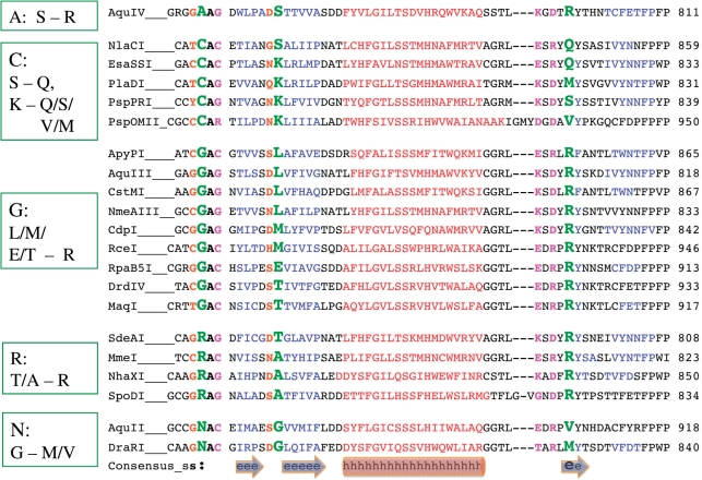
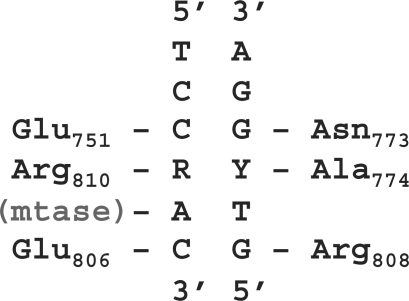
The type II restriction endonucleases are indispensible tools for molecular biology. Although enzymes recognizing nearly 300 unique sequences are known, the ability to engineer enzymes to recognize any sequence of choice would be valuable. However, previous attempts to engineer new recognition specificity have met limited success. Here we report the rational engineering of multiple new type II specificities. We recently identified a family of MmeI-like type II endonucleases that have highly similar protein sequences but different recognition specificity. We identified the amino-acid positions within these enzymes that determine position specific DNA base recognition at three positions within their recognition sequences through correlations between their aligned amino-acid residues and aligned recognition sequences. We then altered the amino acids at the identified positions to those correlated with recognition of a desired new base to create enzymes that recognize and cut at predictable new DNA sequences. The enzymes so altered have similar levels of endonuclease activity compared to the wild-type enzymes. Using simple and predictable mutagenesis in this family it is now possible to create hundreds of unique new type II restriction endonuclease specificities. The findings suggest a simple mechanism for the evolution of new DNA specificity in Nature.
Figures
Similar articles
-
Specificity changes in the evolution of type II restriction endonucleases: a biochemical and bioinformatic analysis of restriction enzymes that recognize unrelated sequences.J Biol Chem. 2005 Feb 11;280(6):4289-98. doi: 10.1074/jbc.M409020200. Epub 2004 Nov 24. J Biol Chem. 2005. PMID: 15563460
-
Catalytic domain of restriction endonuclease BmrI as a cleavage module for engineering endonucleases with novel substrate specificities.Nucleic Acids Res. 2007;35(18):6238-48. doi: 10.1093/nar/gkm665. Epub 2007 Sep 13. Nucleic Acids Res. 2007. PMID: 17855396 Free PMC article.
-
The MmeI family: type II restriction-modification enzymes that employ single-strand modification for host protection.Nucleic Acids Res. 2009 Aug;37(15):5208-21. doi: 10.1093/nar/gkp534. Epub 2009 Jul 3. Nucleic Acids Res. 2009. PMID: 19578066 Free PMC article.
-
Recognition and cleavage of DNA by type-II restriction endonucleases.Eur J Biochem. 1997 May 15;246(1):1-22. doi: 10.1111/j.1432-1033.1997.t01-6-00001.x. Eur J Biochem. 1997. PMID: 9210460 Review.
-
[Type IIE and IIF restriction endonucleases interacting with two recognition sites in DNA].Mol Biol (Mosk). 2004 Sep-Oct;38(5):886-900. Mol Biol (Mosk). 2004. PMID: 15554190 Review. Russian.
Cited by
-
Sequence-specific cleavage of RNA by Type II restriction enzymes.Nucleic Acids Res. 2010 Dec;38(22):8257-68. doi: 10.1093/nar/gkq702. Epub 2010 Aug 11. Nucleic Acids Res. 2010. PMID: 20702422 Free PMC article.
-
Type I restriction enzymes and their relatives.Nucleic Acids Res. 2014 Jan;42(1):20-44. doi: 10.1093/nar/gkt847. Epub 2013 Sep 24. Nucleic Acids Res. 2014. PMID: 24068554 Free PMC article. Review.
-
Crystallization and preliminary crystallographic analysis of the type IIL restriction enzyme MmeI in complex with DNA.Acta Crystallogr Sect F Struct Biol Cryst Commun. 2011 Oct 1;67(Pt 10):1262-5. doi: 10.1107/S1744309111028041. Epub 2011 Sep 30. Acta Crystallogr Sect F Struct Biol Cryst Commun. 2011. PMID: 22102043 Free PMC article.
-
DNA target recognition domains in the Type I restriction and modification systems of Staphylococcus aureus.Nucleic Acids Res. 2017 Apr 7;45(6):3395-3406. doi: 10.1093/nar/gkx067. Nucleic Acids Res. 2017. PMID: 28180279 Free PMC article.
-
Characterization of cleavage intermediate and star sites of RM.Tth111II.Sci Rep. 2014 Jan 23;4:3838. doi: 10.1038/srep03838. Sci Rep. 2014. PMID: 24452415 Free PMC article.
References
-
- Halford S.E. The specificity of the EcoRI restriction endonuclease. Biochem. Soc. Trans. 1980;8:399–400. - PubMed
-
- Heitman J., Ivanenko T., Kiss A. DNA nicks inflicted by restriction endonucleases are repaired by a RecA- and RecB-dependent pathway in Escherichia coli. Mol. Microbiol. 1999;33:1141–1151. - PubMed
-
- Bujnicki J.M. Crystallographic and bioinformatic studies on restriction endonucleases: Inference of evolutionary relationships in the ‘midnight zone' of homology. Curr. Protein Pept. Sci. 2003;4:327–337. - PubMed
Publication types
MeSH terms
Substances
LinkOut - more resources
Full Text Sources
Other Literature Sources
Molecular Biology Databases
Miscellaneous
