. 2009 Aug 28;284(35):23580-91.
doi: 10.1074/jbc.M109.038133. Epub 2009 Jun 30.

Amino acid position-specific contributions to amyloid beta-protein oligomerization

Affiliations

Amino acid position-specific contributions to amyloid beta-protein oligomerization

Samir K Maji et al. J Biol Chem. .

Abstract

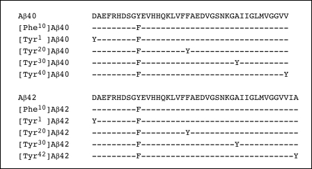
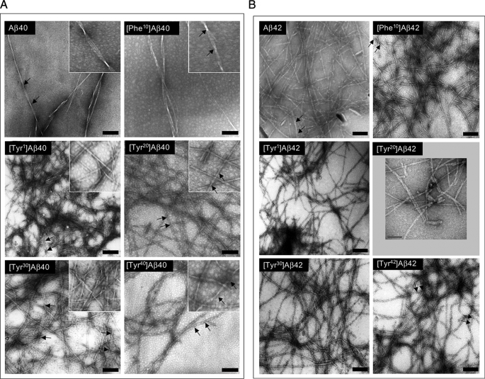
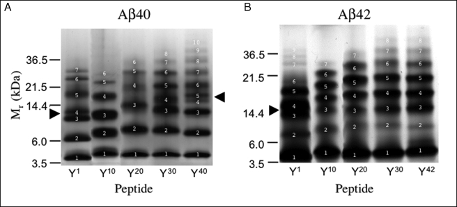
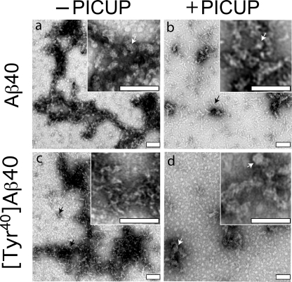
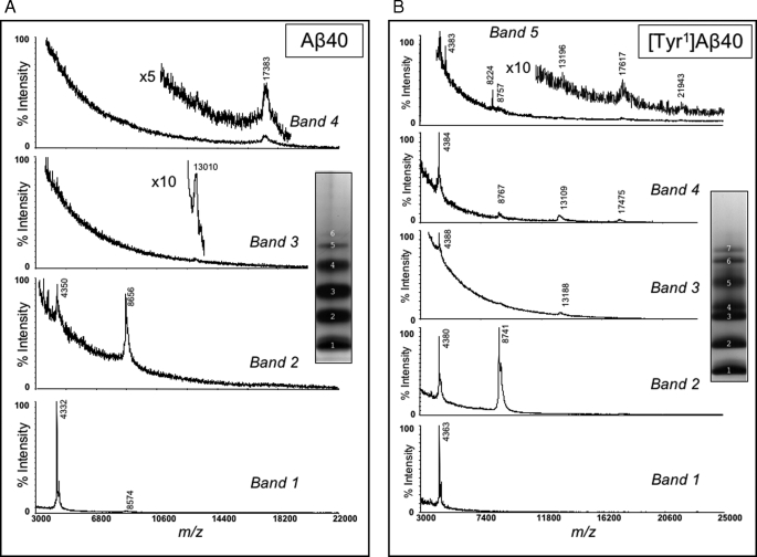
Understanding the structural and assembly dynamics of the amyloid beta-protein (Abeta) has direct relevance to the development of therapeutic agents for Alzheimer disease. To elucidate these dynamics, we combined scanning amino acid substitution with a method for quantitative determination of the Abeta oligomer frequency distribution, photo-induced cross-linking of unmodified proteins (PICUP), to perform "scanning PICUP." Tyr, a reactive group in PICUP, was substituted at position 1, 10, 20, 30, or 40 (for Abeta40) or 42 (for Abeta42). The effects of these substitutions were probed using circular dichroism spectroscopy, thioflavin T binding, electron microscopy, PICUP, and mass spectrometry. All peptides displayed a random coil --> alpha/beta --> beta transition, but substitution-dependent alterations in assembly kinetics and conformer complexity were observed. Tyr(1)-substituted homologues of Abeta40 and Abeta42 assembled the slowest and yielded unusual patterns of oligomer bands in gel electrophoresis experiments, suggesting oligomer compaction had occurred. Consistent with this suggestion was the observation of relatively narrow [Tyr(1)]Abeta40 fibrils. Substitution of Abeta40 at the C terminus decreased the population conformational complexity and substantially extended the highest order of oligomers observed. This latter effect was observed in both Abeta40 and Abeta42 as the Tyr substitution position number increased. The ability of a single substitution (Tyr(1)) to alter Abeta assembly kinetics and the oligomer frequency distribution suggests that the N terminus is not a benign peptide segment, but rather that Abeta conformational dynamics and assembly are affected significantly by the competition between the N and C termini to form a stable complex with the central hydrophobic cluster.


Figures

FIGURE 1.
FIGURE 1.
Primary structure of Aβ peptides. The sequences of wild type Aβ40 and Aβ42 are presented, below which are the sequences of the substituted peptides. Hyphens indicate identical amino acid residues. In peptides in which the Tyr probe was placed at positions other than the native position 10, a Phe group was substituted at position 10. For simplicity, these Aβ homologues are specified only by the position of the Tyr, i.e. [Tyrn]Aβ40/42. The complete peptide specification would include the positions of both the Tyr and Phe residues, e.g. [Tyrn,Phe10]Aβ40/42.
FIGURE 2.
FIGURE 2.
Secondary structure dynamics. A, Aβ40 and homologues were incubated in 10 mm phosphate buffer, pH 7.4, at 22 °C. CD spectra were acquired daily for 14 days. The day on which a spectrum was acquired is indicated by “d.” The spectra shown are the average of six scans each with an averaging time of 5 s. B, Aβ42 and homologues were analyzed in the same manner. Results for both sets of peptides are representative of those obtained in each of four independent experiments.
FIGURE 3.
FIGURE 3.
Morphology of Aβ assemblies. Following peptide assembly, transmission electron microscopy was performed on negatively stained samples as follows: A, Aβ40 and homologues; B, Aβ42 and homologues. The numerous small (<5 nm), translucent background structures visible to various degrees in the panels are not proteinaceous but rather are artifacts of the staining procedure. Protein structures of these sizes are not observed in experiments in which fibril formation is allowed to proceed to completion. Scale bars, 100 nm. Insets in A are higher magnification images of the respective fields. The insets are 133 nm square. Arrows delimit helical pitches discussed in the text.
FIGURE 4.
FIGURE 4.
Oligomer size distributions. A, Aβ40 and homologues were cross-linked using PICUP, and then oligomer frequency distributions were determined by SDS-PAGE followed by silver staining. Molecular masses of protein standards are shown on the left. The gels are representative of each of three independent experiments. B, Aβ42 and homologues analyzed as in A. The arrowheads indicate regions in which band migration differed from that of the corresponding wild type peptide (see text). White numbers specify band numbers.
FIGURE 5.
FIGURE 5.
Morphologic analysis of cross-linked peptides. The morphologies of uncross-linked (−PICUP) (a and c) and cross-linked (+PICUP) (b and d) wild type and Tyr1-substituted Aβ40 peptides, respectively, were determined by EM of negatively stained preparations. Scale bars, 100 nm. The images are representative of those in each of at least three independent experiments. Arrows identify structures discussed in the text.
FIGURE 6.
FIGURE 6.
MALDI-TOF MS analysis of isolated oligomers. Bands produced by Aβ40 (A) and [Tyr1]Aβ40 (B) following PICUP and SDS-PAGE were identified by negative staining of the gels, after which the protein components were eluted and analyzed mass spectrometrically (see “Experimental Procedures”). Normalized ion intensities are presented on the ordinates, and mass-to-charge (m/z) ratios are presented on the abscissas. Band numbers and the locations of specific oligomers within the spectra are indicated. Insets show the actual gel lanes from which the bands were isolated.

References

    1. Hebert L. E., Scherr P. A., Bienias J. L., Bennett D. A., Evans D. A. (2003) Arch. Neurol. 60,1119–1122 - PubMed
    1. Brookmeyer R., Johnson E., Ziegler-Graham K., Arrighi H. M. (2007) Alzheimers Dement. 3,186–191 - PubMed
    1. Kirkitadze M. D., Bitan G., Teplow D. B. (2002) J. Neurosci. Res. 69,567–577 - PubMed
    1. Klein W. L., Stine W. B., Jr., Teplow D. B. (2004) Neurobiol. Aging 25,569–580 - PubMed
    1. Klyubin I., Walsh D. M., Cullen W. K., Fadeeva J. V., Anwyl R., Selkoe D. J., Rowan M. J. (2004) Eur. J. Neurosci. 19,2839–2846 - PubMed

Publication types