. 2009 Oct 8;114(15):3244-54.
doi: 10.1182/blood-2009-04-217620. Epub 2009 Jun 30.

Epigenetic regulation of the alternatively activated macrophage phenotype

Affiliations

Epigenetic regulation of the alternatively activated macrophage phenotype

Makoto Ishii et al. Blood. .

Abstract

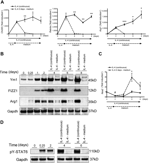
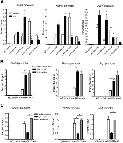
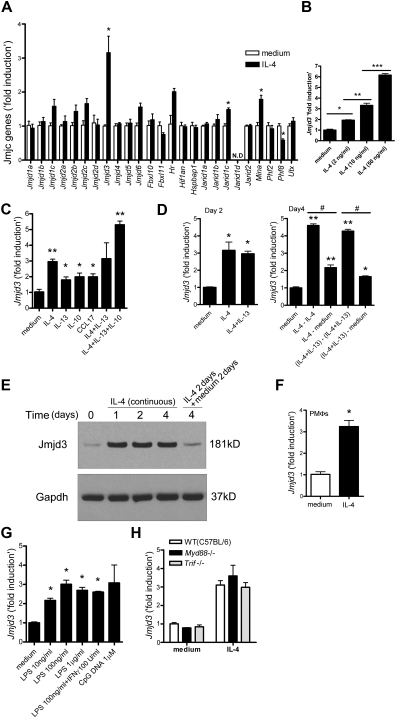
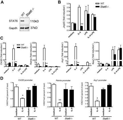
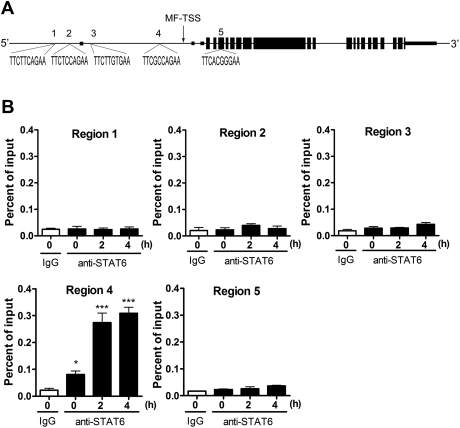
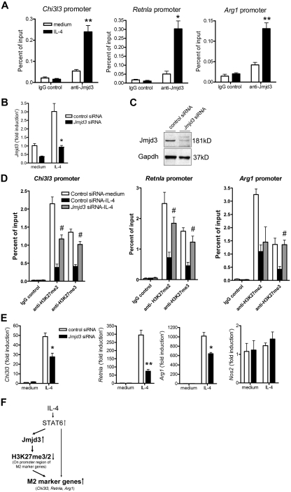
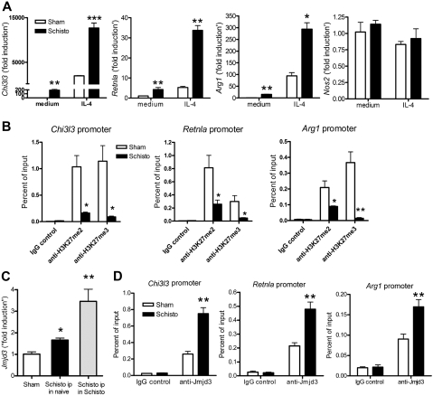
Alternatively activated (M2) macrophages play critical roles in diverse chronic diseases, including parasite infections, cancer, and allergic responses. However, little is known about the acquisition and maintenance of their phenotype. We report that M2-macrophage marker genes are epigenetically regulated by reciprocal changes in histone H3 lysine-4 (H3K4) and histone H3 lysine-27 (H3K27) methylation; and the latter methylation marks are removed by the H3K27 demethylase Jumonji domain containing 3 (Jmjd3). We found that continuous interleukin-4 (IL-4) treatment leads to decreased H3K27 methylation, at the promoter of M2 marker genes, and a concomitant increase in Jmjd3 expression. Furthermore, we demonstrate that IL-4-dependent Jmjd3 expression is mediated by STAT6, a major transcription factor of IL-4-mediated signaling. After IL-4 stimulation, activated STAT6 is increased and binds to consensus sites at the Jmjd3 promoter. Increased Jmjd3 contributes to the decrease of H3K27 dimethylation and trimethylation (H3K27me2/3) marks as well as the transcriptional activation of specific M2 marker genes. The decrease in H3K27me2/3 and increase in Jmjd3 recruitment were confirmed by in vivo studies using a Schistosoma mansoni egg-challenged mouse model, a well-studied system known to support an M2 phenotype. Collectively, these data indicate that chromatin remodeling is mechanistically important in the acquisition of the M2-macrophage phenotype.


Figures

Figure 1
Figure 1
Critical roles of IL-4 on induction and maintenance of alternative activation in BMDMs. BMDMs were incubated with IL-4 for 2 days; then the cells were incubated with (IL-4 continuous group) or without (IL-4 medium group) IL-4 for the indicated time. (A) The mRNA levels of Ym1 (Chi3l3), FIZZ1 (Retnla), and Arginase 1 (Arg1) were analyzed by quantitative RT-PCR. The data shown are “fold induction” relative to that in untreated cells. (B) Expressions of Ym1, FIZZ1, and Arginase 1 (Arg1) were measured by immunoblotting. Gapdh was used as a loading control. (C) The mRNA levels of iNOS (Nos2) were analyzed by quantitative RT-PCR. The data shown are “fold induction” relative to that in untreated cells. #P < .05 compared with the level on day 0. (D) Expression of phosphorylated (pY)-STAT6 was measured by immunoblotting. Gapdh was used as a loading control. Data are representative of 3 independent experiments (A-D) and are expressed as mean ± SEM (A,C). *P < .05, **P < .01, ***P < .001, compared with IL-4 medium group.
Figure 2
Figure 2
Dynamic epigenetic gene regulations in IL-4–induced M2-macrophages. (A) BMDMs were incubated with or without IL-4 for 2 days. ChIP assay was performed on the promoter regions of Chi3l3, Retnla, and Arg1 using indicated antibodies. (B-C) BMDMs were incubated with IL-4 for 2 days and then were cultured with (IL-4 continuous group) or without (IL-4 medium group) IL-4 for an additional 2 days. ChIP assay was performed on the promoter regions of M2 marker genes using antibodies for H3K4me3 (B) and H3K27me3 (C). Untreated cells were used as a baseline control (medium-medium group). Data are representative of 3 to 5 independent experiments and are expressed as mean ± SEM. *P < .05; **P < .01.
Figure 3
Figure 3
Jmjd3 induction in IL-4–treated macrophages. (A) BMDMs were incubated with IL-4 for 6 hours. The mRNA levels of JmjC family proteins were measured by quantitative RT-PCR. N.D. indicates not detected. (B-D) Jmjd3 mRNA levels were measured by quantitative RT-PCR after the following treatments. BMDMs were incubated with IL-4 (B) and indicated cytokines (C) for 6 hours. BMDMs were incubated with indicated cytokines for 2 days (D left). On day 2, cells were washed with phosphate-buffered saline and incubated with or without fresh IL-4, or with or without a cocktail of IL-4 and IL-13 for an additional 2 days as indicated (D right). #P < .05. (E) BMDMs were incubated with IL-4 as indicated. Jmjd3 expression was measured by immunoblotting. Gapdh was used as a loading control. Representative data were shown from 3 independent experiments. (F-H) Jmjd3 mRNA levels were measured by quantitative RT-PCR after the following treatments. Peritoneal macrophages (PMΦs) were incubated with IL-4 for 6 hours (F). BMDMs were incubated with LPS, a cocktail of LPS and IFN-γ, or CpG DNA (in indicated concentration) for 6 hours (G). BMDMs from WT (C57BL/6), Myd88−/−, and Trif−/− mice were incubated with or without IL-4 for 6 hours (H). All data of quantitative RT-PCR shown are “fold induction” relative to that in untreated WT cells. Data are representative of 3 independent experiments and are expressed as mean ± SEM. *P < .05; **P < .01; ***P < .001.
Figure 4
Figure 4
STAT6-dependent Jmjd3 induction in IL-4–induced M2-macrophages. (A) Immunoblotting for STAT6 in BMDMs from WT and Stat6−/− mice. Gapdh was used as a loading control. (B-C) The expression of Jmjd3 (B), Chi3l3, Retnla, Arg1, and Nos2 (C) in WT and Stat6−/− BMDMs was measured by quantitative RT-PCR after the indicated treatments. The data shown are “fold induction” relative to that in untreated WT cells. (D) Anti-H3K27me3 ChIP assay was performed on the promoter regions of M2 marker genes in WT and Stat6−/− BMDMs after IL-4 stimulation. Data are representative of 3 independent experiments and are expressed as mean ± SEM. *P < .05, **P < .01, compared with WT mice group (B-C) or unstimulated (medium alone) group (D).
Figure 5
Figure 5
Direct control of Jmjd3 induction by STAT6. (A) The structure of mouse Jmjd3 gene (NM_001017426). Small boxes represent untranslated regions; larger boxes, coding regions. The putative STAT6-binding sites (regions 1-5) and their sequences are shown. MF-TSS indicates macrophages-transcriptional start site. (B) Anti-STAT6 ChIP assay was performed with primers surrounding STAT6-binding site–containing regions of Jmjd3 gene in BMDMs after IL-4 stimulation. Data are representative of 3 independent experiments and are expressed as mean ± SEM. *P < .05, ***P < .001, compared with IgG control.
Figure 6
Figure 6
M2 marker genes are targets of Jmjd3. (A) Anti-Jmjd3 ChIP assay was performed on the indicated M2 marker promoter regions in BMDMs 48 hours after IL-4 stimulation. (B-E) BMDMs were transfected with Jmjd3 siRNA or control siRNA. The cells were then incubated with IL-4 for 6 hours (B,E) or 24 hours (C), and the expression of indicated genes was measured by quantitative RT-PCR (B,E) or, in the case of Jmjd3, also by immunoblotting (C). Gapdh was used as a loading control. The data shown in panels B and E are “fold induction” relative to that in unstimulated control siRNA-treated cells. Jmjd3 siRNA- or control siRNA-transfected BMDMs were incubated with IL-4 for 48 hours (D). Anti-H3K27me2/3 ChIP assay was performed on the promoter regions of M2 marker genes (D). (F) The schema shows that STAT6-dependent Jmjd3 induction decreases H3K27me2/3 level on M2 marker promoter regions and helps to maintain M2 marker genes in a transcriptionally active state. Data of ChIP assay and quantitative RT-PCR are representative of 3 independent experiments and are expressed as mean ± SEM. *P < .05, **P < .01 compared with unstimulated (medium alone) group (A) or control siRNA group (B,E). #P < .05 compared with control siRNA–IL-4 group (D).
Figure 7
Figure 7
Epigenetic gene regulation of M2-macrophages from S mansoni egg–challenged mice in vivo. Naive mice were intraperitoneally injected with 1.7% sodium chloride solution alone (sham group) or with 5000 S mansoni eggs in 1.7% sodium chloride solution (Schisto group; ie, Schisto ip in naive mice group) for 7 days. (A) The peritoneal macrophages from Schisto and the sham control group were incubated with or without IL-4 for 6 hours. The expression of Chi3l3, Retnla, Arg1, and Nos2 was measured by quantitative RT-PCR. The data shown are “fold induction” relative to that in untreated cells from the sham group. (B) ChIP assay was performed to determine H3K27 methylation status on the promoter regions of Chi3l3, Retnla, and Arg1 in macrophages from both Schisto and sham groups. (C) Naive mice were intraperitoneally injected with 5000 S mansoni eggs (Schisto ip in naive group; ie, Schisto group). In addition, S mansoni–infected mice were intraperitoneally injected with 5000 S mansoni eggs (Schisto ipy in Schisto group). Jmjd3 mRNA levels in peritoneal macrophages from indicated group were measured by quantitative RT-PCR. The data shown are “fold induction” relative to that in cells from the sham group. (D) Anti-Jmjd3 ChIP assay was performed on M2 marker promoter regions in macrophages from Schisto and control groups. Data are represented as mean ± SEM of 8 to 10 mice per group. *P < .05, **P < .01, ***P < .001, compared with sham group.

Comment in

References

    1. Ramalingam TR, Reiman RM, Wynn TA. Exploiting worm and allergy models to understand Th2 cytokine biology. Curr Opin Allergy Clin Immunol. 2005;5:392–398. - PubMed
    1. Gordon S. Alternative activation of macrophages. Nat Rev Immunol. 2003;3:23–35. - PubMed
    1. Mantovani A, Sica A, Locati M. New vistas on macrophage differentiation and activation. Eur J Immunol. 2007;37:14–16. - PubMed
    1. Noel W, Raes G, Hassanzadeh Ghassabeh G, De Baetselier P, Beschin A. Alternatively activated macrophages during parasite infections. Trends Parasitol. 2004;20:126–133. - PubMed
    1. Zhu Z, Zheng T, Homer RJ, et al. Acidic mammalian chitinase in asthmatic Th2 inflammation and IL-13 pathway activation. Science. 2004;304:1678–1682. - PubMed

Publication types

MeSH terms