. 2009 Jan;19(1):3-16.
doi: 10.1055/s-0028-1103121.

Differential diagnosis of jugular foramen lesions

Affiliations

Differential diagnosis of jugular foramen lesions

Thomas J Vogl et al. Skull Base. 2009 Jan.

Abstract

The anatomy of the jugular foramen is complex. It contains the lower cranial nerves and major vascular structures. Tumors that develop within it, or extend into it, provide significant diagnostic and surgical challenges. In this article, we describe the anatomy of the jugular foramen and outline an imaging protocol that can differentiate between lesions, thereby aiding diagnosis and facilitating management.

Keywords: Jugular foramen; computed tomography; magnetic resonance imaging; skull base.


Figures

Figure 1
Figure 1
Three-dimensional volume-rendered reconstruction of the skull base using 64-slice computed tomography (CT) scanner. (A) Inferior view shows oval-shaped jugular fossa (arrow) just behind to the mastoid process. (B) Superior view shows jugular tubercle (white arrowhead) medial to the oval-shaped jugular fossa (black arrow). The impression of the hypoglossal canal (white curved arrow) is inferior and medial to the jugular foramen. The foramen lacerum (asterisk) and foramen ovale (black arrowhead) are also seen.
Figure 2
Figure 2
Computed tomography (CT) and angiographic images of glomus jugular tumor arising in the right jugular foramen. (A) Soft tissue axial postcontrast CT demonstrates a glomus jugular tumor on the right side with small intracranial extension. (B) Bone-windowed axial CT image (after contrast agent administration) shows a large expansile mass in the right jugular fossa protruding into the right hypotympanum. Note the infiltration of the vertical carotid canal, as well as “moth-eaten” borders of the adjacent petrous bone and clivus. (C) Three-dimensional volume rendered image (viewed from above) constructed from spiral CT data (bone-algorithm) illustrating the extent of bone destruction around the right jugular fossa (arrowheads). Compare with the contralateral normal structures. (D) Selective external carotid artery angiogram of the same patient (lateral view) is shown. The characteristically hypervascular tumor with a coarse blush is supplied by the hypertrophic ascending pharyngeal artery and the occipital artery (arrows). Note the early draining vein during the midarterial phase.
Figure 3
Figure 3
Glomus jugular tumor of the left jugular foramen. (A,B) Axial T1-weighted images before (A) and after (B) gadolinium administration and fat-suppression show invasive, slightly hyperintense lesion with marked enhancement in postcontrast images. Note the typical “salt-and-pepper” appearance of the tumor in the postcontrast image. (C) Coronal T2-weighted image demonstrates the tumor's pattern of spread inferiorly and laterally to the middle ear cavity. The tumor did not involve the inner ear.
Figure 4
Figure 4
Schwannoma of the left jugular foramen (JF). (A) Bone-windowed computed tomography (CT) images demonstrate significant enlargement of the JF on the left side. The jugular spine is well delineated. Compare with the “moth-eaten” pattern of Fig. 2B. (B,C) Adjacent axial CT images after contrast agent administration show a fairly well-enhanced lesion (arrow) in the left JF extending extracranially and compressing the neighboring jugular vein (curved arrow). The patient complained of a hoarse voice for 5 months. The schwannoma can be classified as type C. (From Samii M, Babu RP, Tatagiba M, Sepehrnia A. Surgical treatment of jugular foramen schwannomas. J Neurosurg 1995;82:924–932.)
Figure 5
Figure 5
Primary meningioma of the right jugular foramen (JF) (intrinsic lesion). (A) Axial computed tomography (CT) scan with contrast agent shows a large, centrifugally growing, nonhomogeneously enhancing lesion in the right cerebellopontine angle, the JF, and the infratemporal fossa. The ventral borders of the tumor are ill defined, indicating an infiltrative pattern of spread toward the nasopharynx. (B) Coronal CT image with contrast agent and bone-algorithm reconstruction demonstrates a sclerotic jugular fossa on the right side in which the normal architecture has been maintained. The dumbbell-shaped tumor shows a broad dural attachment intracranially, and this provides an important differentiating imaging feature.
Figure 6
Figure 6
Secondary meningioma of the right jugular foramen (JF) (extrinsic lesion). Axial T1-weighted postgadolinium magnetic resonance image shows a small meningioma (arrow) originating from the right JF (arrowhead) and extending medially into the cerebellopontine angle cistern (not shown) with an en plaque growth pattern. The tumor lacks the “salt-and-pepper” pattern of paragangliomas. Note the presence of a “dural tail” sign.
Figure 7
Figure 7
Metastasis of breast cancer in the left jugular foramen. (A) Axial soft tissue algorithm postcontrast computed tomography (CT) demonstrates hypodense tumor located in the left jugular fossa extending into the ventral aspect of the petrous bone. The CT image shows unequivocal tumor invasion of the jugular bulb, which is critical for selection of the surgical approach. Infiltration of the posterior aspect of the longus colli muscle is probable. (B) The corresponding bone-windowed image clearly shows demineralization of the adjacent petrous apex, which has irregular borders.
Figure 8
Figure 8
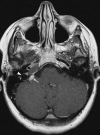
Magnetic resonance imaging (MRI) of retrograde perineural spread from squamous cell carcinoma of the tongue (not shown). Postgadolinium, fat-suppressed T1-weighted MRI shows enlargement and obliteration of the left jugular fossa (arrow) with a moderately enhanced soft tissue mass. The adjacent osseous structures appear diffusely infiltrated, with partial loss of the cortex. Note the perineural spread in the hypoglossal canal (curved arrow), which was responsible for fatty infiltration and atrophy of the left genioglossus and hyoglossus muscles (not shown). Direct encroachment of the medial pterygoid and masseter muscles as well as the parapharyngeal space fat and the pharyngeal constrictor muscle is unequivocal. The MRI findings of the skull base were crucial findings and aided selection of the most appropriate treatment for the patient.
Figure 9
Figure 9
Illustrative example of pseudomass of the left jugular foramen (JF). (A) Axial T1-weighted postcontrast image shows an enlarged JF on the left side with flow-related vivid enhancement. Note the smooth, perfectly rounded outline of the hyperintensity filling the jugular bulb. (B) Three-dimensional time-of-flight venous angiography indicates a high jugular bulb on the left side. Note altered flow of the left transverse sinus, sigmoid sinus, and jugular vein, which accounts for the misdiagnosis.
Figure 10
Figure 10
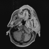
Rhabdomyosarcoma in the right infratemporal fossa. (A) Nonenhanced coronal T1-weighted magnetic resonance imaging (MR) shows a well-circumscribed muscle isointense mass in the right infratemporal fossa. The mass extends into the right jugular foramen (JF) (arrow). Note the marked bilateral cervical lymphadenopathy. (B) Enhanced coronal T1-weighted MRI shows the predominately hyperintense mass extending into the JF (arrow).

References

    1. Tekdemir I, Tuccar E, Aslan A, et al. The jugular foramen: a comparative radioanatomic study. Surg Neurol. 1998;50:557–562. - PubMed
    1. Lang J, Schreiber T. Über form und lage des foramen jugular (fossa jugularis), des canalis caroticus und des foramen stylomastoideum sowie deren postnatale lageveränderungen. HNO. 1983;31:80–87. - PubMed
    1. Ayeni S A, Ohata K, Tanaka K, Hakuba A. The microsurgical anatomy of jugular foramen. J Neurosurg. 1995;83:903–909. - PubMed
    1. Dodo Y. Observations on the bony bridging of the jugular foramen in man. J Anat. 1986;144:153–165. - PMC - PubMed
    1. Shapiro R. Compartmentation of the jugular foramen. J Neurosurg. 1972;36:340–343. - PubMed