

The petro-occipital trans-sigmoid approach for lesions of the jugular foramen
- PMID: 19568342
- PMCID: PMC2637570
- DOI: 10.1055/s-0028-1103127
The petro-occipital trans-sigmoid approach for lesions of the jugular foramen
Abstract
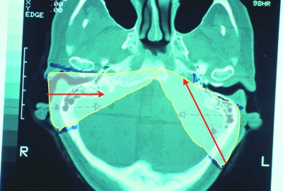
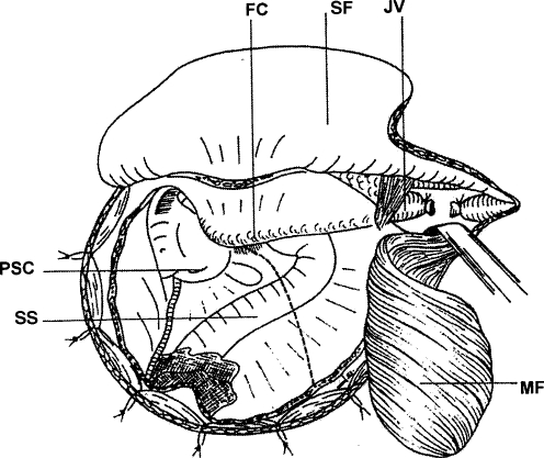
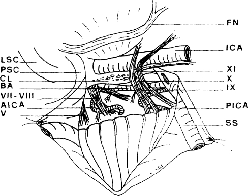
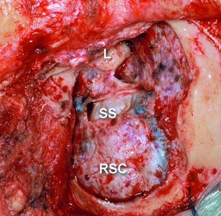
This study's goals were twofold: (1) to analyze the author's experience with the petro-occipital trans-sigmoid (POTS) approach for the resection of tumors arising in or adjacent to the jugular foramen, and (2) to define the anatomical sites exposed by this approach. A retrospective review was conducted of 61 patients with jugular fossa tumors that included lower cranial nerve schwannomas, paragangliomas, meningiomas, chordomas, cholesteatomas, and other benign or low-grade malignant tumors. Outcome measures were mortality, morbidity, and long-term outcomes. No deaths were found in this study. The major morbidity was deficits of the glossopharyngeal, vagus, and accessory nerves. Hearing and facial nerve function were largely preserved. The resections were undertaken as single-stage procedures regardless of whether the tumor was entirely extradural or both intra- and extradural. None of the patients had central nervous system complications. Good outcomes were achieved for schwannomas, meningiomas, chondrosarcomas, and papillary adenoma. Chordomas tended to recur, and only class C1 paragangliomas could be removed using this approach. The study found that the POTS approach should be considered the approach of choice for many tumors in the region of the jugular foramen, particularly schwannomas. It is not suitable for the resection of malignant tumors and most paragangliomas because it offers limited access to the skull base between the jugular fossa and carotid canal.
Keywords: Jugular foramen; petro-occipital trans-sigmoid approach; surgical procedures; tumors of jugular foramen.
Figures
References
-
- Mazzoni A. Jugulo-petrosectomy. Arch Ital Otol Rinol Laringol. 1974;2:20–25.
-
- Gruenert L. Die operative Ausraumung des Bulbus Venae Jugularis (Bulbusoperation) Arch Ohrenheilk. 1884;36:71–77.
-
- Capps F CW. Glomus jugulare tumors of the middle ear. J Laryngol Otol. 1952;66:302–314. - PubMed
-
- Gaillard J, Rebattu J P, Morgan A, Guy F. Note de technique sur la chirurgie des tumeurs glomiques tympano-jugulaires: la dèroutation du nerve facial. J Fr Otorhinolarangol. 1960;9:969–980.
