. 2009 Jul;11(7):637-50.
doi: 10.1593/neo.81686.

Gemcitabine-based chemogene therapy for pancreatic cancer using Ad-dCK::UMK GDEPT and TS/RR siRNA strategies

Affiliations

Gemcitabine-based chemogene therapy for pancreatic cancer using Ad-dCK::UMK GDEPT and TS/RR siRNA strategies

Soukaina Réjiba et al. Neoplasia. 2009 Jul.

Abstract

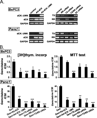
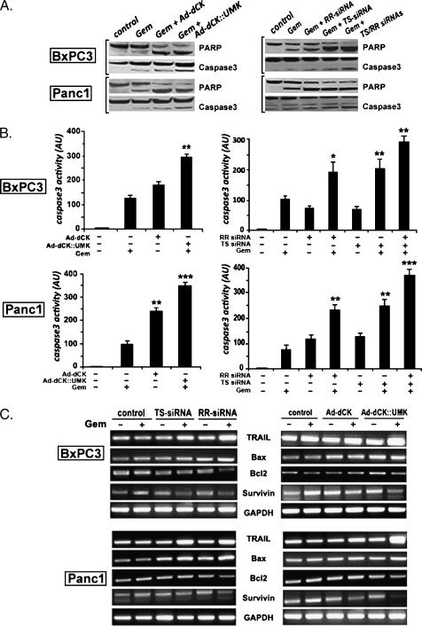
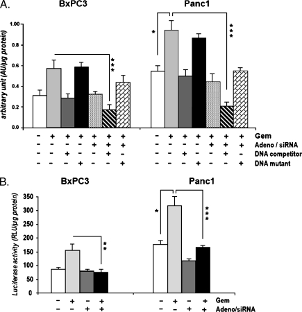
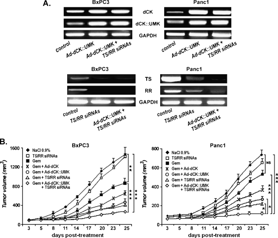
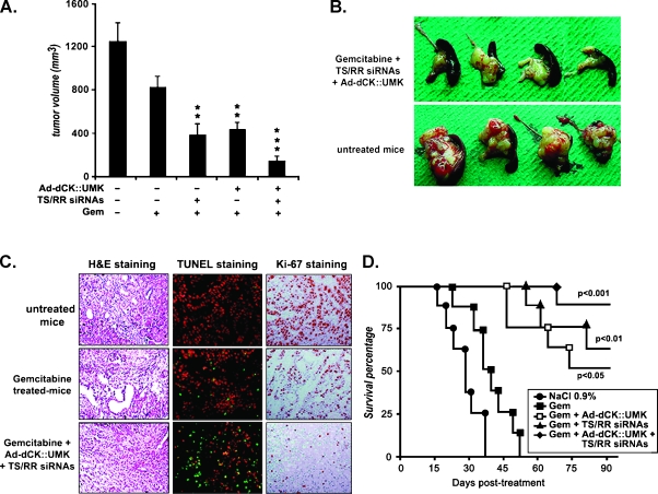
Gemcitabine is a first-line agent for advanced pancreatic cancer therapy. However, its efficacy is often limited by its poor intracellular metabolism and chemoresistance. To exert its antitumor activity, gemcitabine requires to be converted to its active triphosphate form. Thus, our aim was to improve gemcitabine activation using gene-directed enzyme prodrug therapy based on gemcitabine association with the deoxycytidine kinase::uridine monophosphate kinase fusion gene (dCK::UMK) and small interference RNA directed against ribonucleotide reductase (RRM2) and thymidylate synthase (TS). In vitro, cytotoxicity was assessed by 3-[4,5-dimethylthiazol-2-yl]-3,5-diphenyl tetrazolium bromide and [(3)H]thymidine assays. Apoptosis-related gene expression and activity were analyzed by reverse transcription-polymerase chain reaction, Western blot, and ELISA. For in vivo studies, the treatment efficacy was evaluated on subcutaneous and orthotopic pancreatic tumor models. Our data indicated that cell exposure to gemcitabine induced a down-regulation of dCK expression and up-regulation of TS and RR expression in Panc1-resistant cells when compared with BxPc3- and HA-hpc2-sensitive cells. The combination of TS/RRM2 small interference RNA with Ad-dCK::UMK induced a 40-fold decrease of gemcitabine IC(50) in Panc1 cells. This strong sensitization was associated to apoptosis induction with a remarkable increase in TRAIL expression and a diminution of gemcitabine-induced nuclear factor-kappaB activity. In vivo, the gemcitabine-based tritherapy strongly reduced tumor volumes and significantly prolonged mice survival. Moreover, we observed an obvious increase of apoptosis and decrease of cell proliferation in tumors receiving the tritherapy regimens. Together, these findings suggest that simultaneous TS/RRM2-gene silencing and dCK::UMK gene overexpression markedly improved gemcitabine's therapeutic activity. Clearly, this combined strategy warrants further investigation.


Figures

Figure 1
Figure 1
Pancreatic tumor cells sensitivity to gemcitabine. (A) Gemcitabine cytotoxicity. Panc1, BxPc3, and HA-hpc2 cells were treated for 72 hours with increasing concentrations of gemcitabine (0.01–100 µM). Cell viability was measured using MTT test and was presented as a percentage of untreated cell control values. Each curve is representative of four experiments performed in triplicate. (B) Expression of the hENT1 nucleoside transporter. Cells were untreated or treated with gemcitabine for 48 hours, and RNA was extracted and submitted to RT-PCR analysis using specific primers for hENT1 and GAPDH housekeeping gene. (C) Role of hENT1 in gemcitabine transport. BxPc3 and Panc1 cells were treated simultaneously with gemcitabine and the hENT1 inhibitor NBTI (10 and 100 nM). Three days later, cell survival was evaluated by MTT assays. (D) Expression of dCK, RR, and TS. Tumor cells were untreated or treated with gemcitabine at IC50 concentrations, and mRNA levels were evaluated 48 hours later using semiquantitative RT-PCR with specific primers.
Figure 2
Figure 2
Combination of gemcitabine with Ad-dCK::UMK or TS/RRM2 siRNA improves gemcitabine cytotoxic apoptotic effect. (A) Efficacy of recombinant adenovirus transduction and siRNA transfection. BxPc3 and Panc1 cells were either infected with adenoviruses expressing dCK or dCK::UMK or transfected with TS and/or RRM2 siRNA. The RT-PCR analysis was carried out after a 48-hour treatment period on the total RNA using specific primers. dCK, dCK::UMK, RR, and TS expressions were compared with untreated control cells. The nonspecific scramble (Sc) siRNA was used as a negative control for siRNA. (B) Effect of Ad-dCK::UMK and TS/RRM2 siRNA on gemcitabine cytotoxicity. Cells were treated as described above and further received increasing concentrations of gemcitabine for 72 hours. In [3H]thymidine incorporation and MTT assays, results of gemcitabine combined treatments are relative to Ad-dCK::UMK, TS, and/or RR siRNA treatments. The average percentage values obtained in cells treated with gemcitabine alone are relative to those of untreated cells. Values represent the mean of four experiments performed in triplicate. Asterisks indicate significant difference (*P < .05, **P < .01, and ***P < .001) observed after the various combined treatments when compared with gemcitabine alone.
Figure 3
Figure 3
Combination of gemcitabine with Ad-dCK::UMK or TS/RRM2 siRNA improves gemcitabine cytotoxic apoptotic effect. (A) Enhancement of PARP and caspase-3 activation. To assess PARP and caspase-3 activation, Western blot analysis experiments were performed on cell lysates obtained from cells treated with either Ad-dCK::UMK or TS/RRM2 siRNA and further treated with gemcitabine (IC50 doses) during 48 hours. The upper panels represent the 116-kDa native form and the 85 kDa activated form of PARP. The lower panels illustrate the native and the cleaved forms of caspase-3 (17 and 20 kDa). (B) Quantitative measurement of caspase-3 activity. Caspase-3 activation was also evaluated by colorimetric assays. Data are representative of three independent experiments. Asterisks indicate significant difference (**P < .01 and ***P < .001) observed after the various treatments when compared with gemcitabine alone. (C) Expression study of different apoptotic genes. Treated cells were submitted to gemcitabine treatment for 48 hours and then used for TRAIL, Bax, Survivin, and Bcl2 mRNA level measurement using RT-PCR analysis. GAPDH was used as an internal control.
Figure 4
Figure 4
Maximal optimization of gemcitabine efficacy is obtained by combination with Ad-dCK::UMK and TS/RR siRNA. (A) Cytotoxic effect of Gemcitabine combination with Ad-dCK::UMK and TS/RR siRNA. Cell viability was determined on Ad-dCK::UMK-infected and/or TS/RR siRNA-transfected cells by MTT assay after 72 hours of treatment with increasing concentrations of gemcitabine. The average percentages of the MTT value obtained in gemcitabine-treated cells are relative to those of untreated cells. Concerning the gemcitabine combined treatments, results represent relative value of gemcitabine-combined treatments to Ad-dCK::UMK, TS/RR siRNA, or Ad-dCK::UMK plus TS/RR siRNA treatments. Data are the result of three independent experiments. Asterisks indicate significant difference (*P < .05, **P < .01, and ***P < .001) observed after the various treatments when compared with gemcitabine alone. (B) Hoechst staining. Cells were treated as indicated in the Materials and Methods section. Apoptotic cells were visualized using fluorescent microscope. (C) Caspase-3 activation. Activity of caspase-3 was studied by Western blot analysis and colorimetric activity assay on cells having received Ad-dCK::UMK plus TS/RRM2 siRNA and further treated for 48 hours with gemcitabine at IC50 doses. Asterisks indicate significant difference (**P < .01 and ***P < .001) observed after cell treatment with gemcitabine associated to both Ad-dCK::UMK and TS/RR siRNA treatments compared with cells treated with gemcitabine plus Ad-dCK::UMK or gemcitabine plus TS/RRM2 siRNA. (D) Expression of apoptotic mediators. In the same experiment, TRAIL, Bax, Bcl2, and Survivin apoptotic gene expressions were evaluated by RT-PCR experiments using their respective specific primers. The expression of GAPDH is shown as an internal control.
Figure 5
Figure 5
Role of NF-κB in tumor cell sensitivity to gemcitabine alone or in combination with Ad-dCK::UMK and TS/RRM2 siRNA. BxPc3 and Panc1 tumor cells were cotreated with Ad-dCK::UMK and TS/RR siRNA. One day later, cells further received 2 and 40 µM of gemcitabine, respectively, and NF-κB activation was studied after 48 hours. (A) NoShift NF-κB colorimetric assay. Cells were harvested, and NF-κB activity was evaluated according to the manufacturer's recommendations. (B) NF-κB luciferase gene reporter assay. Herein, cells were primary transfected with a plasmid-expressing luciferase under control of an NF-κB response element. Luciferase assays were then performed according to the manufacturer's protocol. All experiments were performed in triplicate and repeated at least three times. Asterisks indicate significant difference (**P < .01 and ***P < .001) observed in cells treated with the tritherapy gemcitabine plus Ad-dCK::UMK plus TS/RR siRNA compared with cells treated with gemcitabine alone. *P < .05 indicates the significant difference observed between gemcitabine-treated Panc1 cells relative to untreated Panc1 cells.
Figure 6
Figure 6
Overexpression of dCK::UMK and down-regulation of TS/RR improved gemcitabine's therapeutic effect in subcutaneous pancreatic tumor models. Nude mice were subcutaneously inoculated with BxPc3 or Panc1 cells, and when their tumor size reached 100 mm3, they received intratumor injection of Ad-dCK::UMK (1 x 10⁹ PFU) ± TS/RR siRNA (100 µg). Gemcitabine (15 mg/kg body weight) was administrated intraperitoneally. Control group was treated with 0.9% NaCl. (A) Ad-dCK::UMK and TS/RR siRNA functionality. Tumors were recovered from two mice per group, and RT-PCR experiments were carried out to demonstrate the reduction of endogenous TS and RR expression and the overexpression of dCK and dCK::UMK after siRNA and Ad-dCK::UMK treatments, respectively. GAPDH was used as an internal control. (B) Tumor growth. Tumor growth evolution of BxPc3 and Panc1 tumors was followed up by tumor volume measurement two times weekly. NS indicates not significant. **P < .01 and ***P < .001 versus control.
Figure 7
Figure 7
Inhibition of orthotopic tumor formation and increased survival of tumor-bearing mice after combined gemcitabine-based treatment. Thirty days after the beginning of treatment, six mice from each group (n = 14) were killed; their pancreatic tumors were recovered. (A) Tumor growth. Recovered biopsies were measured in three dimensions. Asterisks indicate significant difference (**P < .01 and ***P < .001) observed after gemcitabine combined treatments compared with gemcitabine alone. (B) Macroscopic findings of pancreas. Photographs represent excised pancreatic tumor from untreated mice and those receiving tritherapy protocol. (C) Representative immunohistochemistry examination. Biopsies were subsequently submitted to hematoxylin-eosin staining (left panels), TUNEL staining (middle panels), and Ki-67 staining (right panels). (D) Kaplan-Meier survival curve. Remaining mice were followed for survival curve determination. Results were analyzed statistically by log-rank test. Asterisks indicate significant difference (*P < .5, **P < .01, and ***P < .001) observed after gemcitabine combined treatments compared with gemcitabine alone.

References

    1. Yeo TP, Hruban RH, Leach SD, Wilentz RE, Sohn TA, Kern SE, Iacobuzio-Donahue CA, Maitra A, Goggins M, Canto MI, et al. Pancreatic cancer. Curr Probl Cancer. 2002;26:176–275. - PubMed
    1. Kollmannsberger C, Peters HD, Fink U. Chemotherapy in advanced pancreatic adenocarcinoma. Cancer Treat Rev. 1998;24:133–156. - PubMed
    1. Burris HA, Moore MJ, Andersen J, Green MR, Rothenberg ML, Modiano MR, Cripps MC, Portenoy RK, Storniolo AM, Tarassoff P, et al. Improvements in survival and clinical benefit with gemcitabine as first-line therapy for patients with advanced pancreatic cancer: a randomized-trial. J Clin Oncol. 1997;15:2403–2413. - PubMed
    1. Burris H, Storniolo AM. Assessing clinical benefit in the treatment of pancreas cancer: gemcitabine compared to 5-fluorouracil. Eur J Cancer. 1997;33(Suppl):18–22. - PubMed
    1. Rothenberg ML, Moore MJ, Cripps MC, Andersen JS, Portenoy RK, Burris HA, III, Green MR, Tarassoff PG, Brown TD, Casper ES, et al. A phase II trial of gemcitabine in patients with 5-FU-refractory pancreas cancer. Ann Oncol. 1996;7:347–353. - PubMed

Publication types

MeSH terms