. 2009 Jul 1;4(7):e6123.
doi: 10.1371/journal.pone.0006123.

Intracellular localization and conformational state of transglutaminase 2: implications for cell death

Affiliations

Intracellular localization and conformational state of transglutaminase 2: implications for cell death

Soner Gundemir et al. PLoS One. .

Abstract

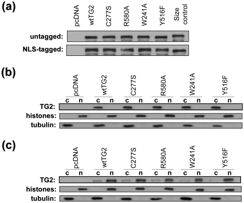
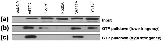
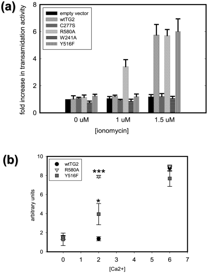
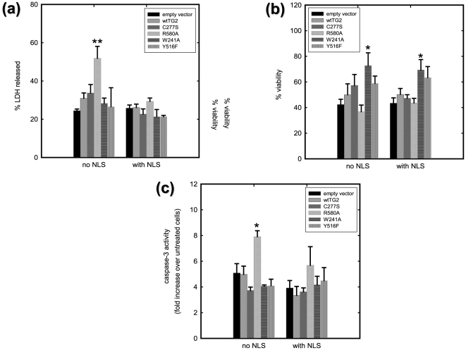
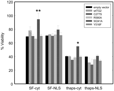
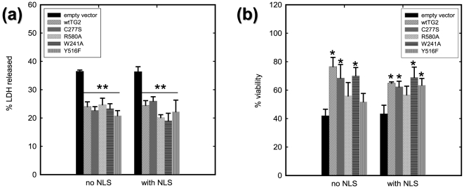
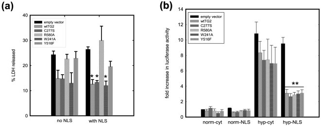
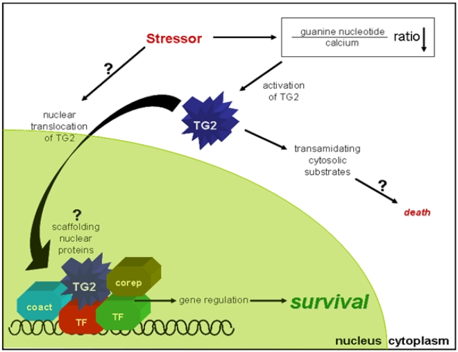
Transglutaminase 2 (TG2) is a multifunctional enzyme that has guanine nucleotide binding and GTP hydrolyzing activity in addition to its transamidating function. Studies show that TG2 is a player in mediating cell death processes. However, there is far from a consensus about the role of this enzyme in cell death processes as it appears to be dependent upon the cell type, stimuli, subcellular localization and conformational state of the enzyme. The purpose of this study was to dissect the role of TG2 in the cell death processes. To this end, we created and characterized 4 distinct point mutants of TG2, each of which differs from the wild type by its conformation or by lacking an important function. We also prepared these mutants as nuclear targeted proteins. By overexpressing mutant or wild type forms of TG2 in HEK 293 cells, we investigated the modulatory role of the protein in the cell death process in response to three stressors: thapsigargin, hyperosmotic stress and oxygen/glucose deprivation (OGD). All of the TG2 constructs, except the R580A mutant (which cannot bind guanine nucleotides and is therefore more prone to exhibit transamidating activity), either did not significantly affect the cell death processes or were protective. However in the case of the R580A mutant, cell death in response to high thapsigargin concentrations, was significantly increased. Intriguingly, nuclear localization of R580A-TG2 was sufficient to counteract the pro-death role of cytoplasmic R580A-TG2. In addition, nuclear localization of TG2 significantly facilitated its protective role against OGD. Our data support the hypothesis that the transamidation activity of TG2, which is mostly quiescent except in extreme stress conditions, is necessary for its pro-death role. In addition, nuclear localization of TG2 generally plays a key role in its protective function against cell death processes, either counteracting the detrimental effect or strengthening the protective role of the protein.


Conflict of interest statement

Competing Interests: The authors have declared that no competing interests exist.

Figures

Figure 1
Figure 1. Expression and Subcellular Localization of TG2 constructs.
Representative immunoblots of cell lysates from HEK 293A cells which were transiently transfected with TG2 constructs. (a) Expression of TG constructs. Lysates were blotted for TG2. untagged: TG2s without an NLS tag, except size control (which is NLS-tagged wild type). NLS-tagged: TG2s with NLS tag, except lane size control (which is untagged wild type). (b) Subcellular localization of untagged TG2 variants. Twenty four hours posttransfection cells were separated into nuclear and cytosolic fractions and were blotted for TG2, histones and tubulin proteins. Histone (nuclear marker) and tubulin (cytosolic marker) blots show the purity of the fractions. TG2 blot shows that untagged TG2 variants are localized almost exclusively to the cytosol and none of the mutations has a significant effect on the localization of TG2. (c) Subcellular localization of NLS-tagged TG2 variants. Twenty four hours posttransfection cells were separated into nuclear and cytosolic fractions and were blotted for TG2, histones and tubulin proteins. TG2 blot shows that more than 80% of the total amount of NLS-tagged TG2 variants are localized to the nucleus and none of the mutations has a significant effect on the localization of NLS-TG2. 10 µg of protein was loaded in each well.
Figure 2
Figure 2. GTP Binding of TG2 Mutants.
Immunoblot showing GTP binding of different TG2 constructs. (a) expression levels of TG2 constructs. 10 µg of protein was loaded in each well. Pulldown experiments were performed with GTP agarose using low (b) and high (c) stringency conditions to evaluate the strength of binding between TG2 and GTP.
Figure 3
Figure 3. Transamidating activity of TG2 variants.
Graphs showing in situ (a) and in vitro (b) transamidating activities of different TG2 mutants as a function of ionomycin concentration (µM) in the cell culture medium (a) or calcium ion added (mM) to the reaction mixture (b) (N = 3). Results are shown as mean+/−SE *p<0.05, **p<0.01, ***p<0.005.
Figure 4
Figure 4. TG2 can either protect against or facilitate thapsigargin induced cell death in HEK 293A cells depending on its conformation and its localization.
(a) LDH release after 9 h of 15 µM thapsigargin treatment. LDH release is significantly increased in HEK 293A cells which express R580A-TG2 without an NLS tag. (N = 3). (b) Cell viability determined by the resazurin assay after 10 h of 15 µM thapsigargin treatment. Metabolic activity is significantly increased in HEK 293A cell which express W241A-TG2 with or without an NLS tag. (N = 4) (c) Caspase-3 activity after 6 h of 15 µM thapsigargin treatment. Caspase-3 activity is significantly increased in HEK 293A cells which express R580A without an NLS tag. (N = 3). Results are shown as mean+/−SE *p<0.05, **p<0.01.
Figure 5
Figure 5. Cytosolic W241A-TG2 protects against serum starvation and thapsigargin-induced toxicity in HEK 293TN cells.
Cell viability determined by the resazurin assay after 24 h of serum starvation and serum starvation combined with 2.5 µM thapsigargin treatment. Cytosolic W241A-TG2 significantly improves metabolic activity of HEK 293TN cell both after serum starvation alone and serum starvation combined with thapsigargin treatment. (N = 3) Results are shown as mean+/−SE *p<0.05, **p<0.01.
Figure 6
Figure 6. TG2 protects HEK 293A cells from hyperosmotic stress, independent of its localization and conformation.
(a) LDH release after 10 h of 1.0 M sorbitol treatment. LDH release is significantly decreased in HEK 293A cells which express TG2 regardless of the conformation state or its subcellular localization (N = 5). (b) Cell viability determined by the resazurin assay after 6 h of 1.0 M sorbitol treatment. Hyperosmotic stress-induced decreases in metabolic activity are significantly attenuated by TG2 constructs except for untargetted R580A-TG2, untargetted Y516F-TG2 and nuclear targetted R580A-TG2 (N = 4). Results are shown as mean+/-SE *p<0.05, **p<0.01.
Figure 7
Figure 7. Nuclear targeted and transamidating inactive TG2 protects HEK 293A cells from oxygen/glucose deprivation triggered cell death independent of HIF signaling.
(a) LDH release after 16 h of oxygen/glucose deprivation (OGD). LDH release is significantly decreased in HEK 293A cells which express wild type-TG2, C277S-TG2 or W241A-TG2 with an NLS tag upon oxygen/glucose deprivation. (N = 5) (b) HRE luciferase activity after 16 h of 0.1% oxygen treatment. All nuclear targeted TG2 variants significantly decreased hypoxia responsive gene transcription whereas none of the untargeted TG2 variants had a significant effect. Results are shown as mean+/−SE *p<0.05, **p<0.01.
Figure 8
Figure 8. Hypothetical model of the role of TG2 in cell death/survival paradigm.
Depending on the stressor type and intensity, TG2 can facilitate or counteract cell death process. Some stressors, such as OGD, trigger nuclear translocation of TG2 through an unknown mechanism, where TG2 mainly promotes survival processes possibly through intervening transcriptional machinery by scaffolding certain transcription factors (TF), co-activators (coact) or co-repressors (corep). Conversely, some stressors dramatically decrease the guanine nucleotide to calcium ratio and elevate transamidating activity of TG2. The increase in the transamidating activity generally means that the death decision has been made and the transamidating function of TG2 helps to execute the death process.

References

    1. Griffin M, Casadio R, Bergamini CM. Transglutaminases: nature's biological glues. Biochem J. 2002;368:377–396. - PMC - PubMed
    1. Chen JS, Mehta K. Tissue transglutaminase: an enzyme with a split personality. Int J Biochem Cell Biol. 1999;31:817–836. - PubMed
    1. Begg GE, Carrington L, Stokes PH, Matthews JM, Wouters MA, et al. Mechanism of allosteric regulation of transglutaminase 2 by GTP. Proc Natl Acad Sci U S A. 2006;103:19683–19688. - PMC - PubMed
    1. Nakaoka H, Perez DM, Baek KJ, Das T, Husain A, et al. Gh: a GTP-binding protein with transglutaminase activity and receptor signaling function. Science. 1994;264:1593–1596. - PubMed
    1. Hasegawa G, Suwa M, Ichikawa Y, Ohtsuka T, Kumagai S, et al. A novel function of tissue-type transglutaminase: protein disulphide isomerase. Biochem J. 2003;373:793–803. - PMC - PubMed

Publication types