Review
. 2009 Jul;27(1):E4.
doi: 10.3171/2009.4.FOCUS0979.

Evolution of brain-computer interfaces: going beyond classic motor physiology

Affiliations
Review

Evolution of brain-computer interfaces: going beyond classic motor physiology

Eric C Leuthardt et al. Neurosurg Focus. 2009 Jul.

Abstract

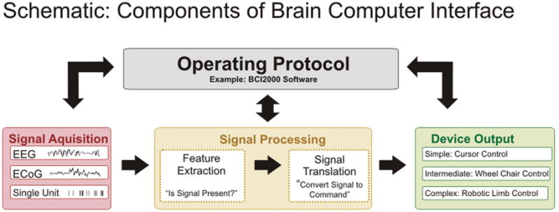
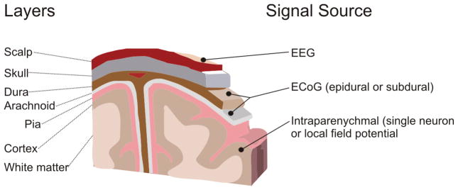
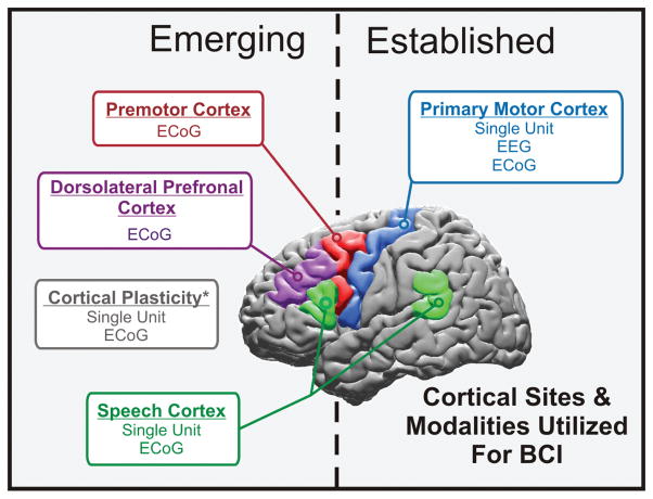
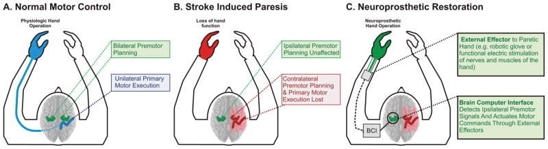
The notion that a computer can decode brain signals to infer the intentions of a human and then enact those intentions directly through a machine is becoming a realistic technical possibility. These types of devices are known as brain-computer interfaces (BCIs). The evolution of these neuroprosthetic technologies could have significant implications for patients with motor disabilities by enhancing their ability to interact and communicate with their environment. The cortical physiology most investigated and used for device control has been brain signals from the primary motor cortex. To date, this classic motor physiology has been an effective substrate for demonstrating the potential efficacy of BCI-based control. However, emerging research now stands to further enhance our understanding of the cortical physiology underpinning human intent and provide further signals for more complex brain-derived control. In this review, the authors report the current status of BCIs and detail the emerging research trends that stand to augment clinical applications in the future.


Figures

Fig. 1
Fig. 1
Schematic showing essential features and components of a BCI. There are 4 essential elements to the practical functioning of a BCI platform: 1) signal acquisition, the BCI system’s recorded brain signal or information input; 2) signal processing, the conversion of raw information into a useful device command; 3) device output, the overt command or control functions administered by the BCI system; and 4) operating protocol, the manner in which the system is turned on and off and the way in which the user or a technical assistant adjusts the parameters of the previous 3 steps in converting intentions to machine commands. All of these elements act in concert to manifest the user’s intention to his or her environment.
Fig. 2
Fig. 2
Drawing depicting the signals for BCI and their locations relative to the brain. Three general categories of signals are used for BCI applications.
Fig. 3
Fig. 3
Schematic showing a summary of the cortical sites and modalities used for BCI. The 3 fundamental signal modalities currently being explored include EEG, ECoG, and single-unit systems that record action potential firing from single neurons.
Fig. 4
Fig. 4
Diagram showing ipsilateral BCI for hemispheric stroke. In the normal physiological scenario (A) motor planning is represented in both hemispheres and motor execution is accomplished by the contralateral M1. In the setting of stroke (B), contralateral primary motor and motor-associated cortex is lost. Premotor cortex ipsilateral to the affected limb is left unaffected. There is an increase in ipsilateral premotor activity following hemispheric stroke. Thus, in the scenario of hemispheric stroke with contralesional premotor upregulation, a BCI can provide a unique opportunity to aid in actuating the nascent premotor commands. A BCI detects the brain signals associated with these premotor commands (C) and converts these signals into machine commands that can control a robotic assist device, which would in turn allow improved hand function (that is, a robotic glove that opens and closes the hand). The BCI allows the ipsilateral premotor cortex to bypass the physiological bottleneck determined by the injured contralateral M1 and small, variable percentage of ipsilateral uncrossed motor fibers.
Fig. 5
Fig. 5
Utilizing cortical plasticity for device control. To achieve 2D control, the amplitude of the signal between 65–100 Hz from one epidural ECoG electrode was used as the control for the horizontal velocity of the cursor, and a separate electrode was used for the vertical velocity of the cursor. The 2 sites were ~ 1 cm apart. For the monkey to improve his performance in a circle-drawing task, it must gain independent control of the 2 signals being used for control. For a perfectly drawn circle, the overall correlation between the 2 signals will be 0. This decorrelation could be done either indiscriminately across all frequencies or only within the frequency band being used for control. To examine what actually occurred during the experiment, the power spectrum was calculated for the 2 recorded signals in 300-msec nonoverlapping time bins. The correlation between the powers at each given frequency for the 2 different channels was then calculated for all points in time. Graph showing that the correlation between the recording sites decreased across most frequencies but most dramatically between 65–100 Hz. Therefore, these data clearly show that through biofeedback, motor cortex is quite adaptable to learning and improving BCI control.

References

    1. Abidian MR, Martin DC. Experimental and theoretical characterization of implantable neural microelectrodes modified with conducting polymer nanotubes. Biomaterials. 2008;29:1273–1283. - PMC - PubMed
    1. Andersen RA, Burdick JW, Musallam S, Pesaran B, Cham JG. Cognitive neural prosthetics. Trends Cogn Sci. 2004;8:486–493. - PubMed
    1. Anderson N, Sharma M, Wisneski K, Blakely T, Shalk G, Moran D, et al. Directional encoding in motor cortex with ipsilateral EcoG activity. Washington, DC: Society for Neuroscience Annual Meeting; 2008.
    1. Billingsley-Marshall RL, Clear T, Mencl WE, Simos PG, Swank PR, Men D, et al. A comparison of functional MRI and magnetoencephalography for receptive language mapping. J Neurosci Methods. 2007;161:306–313. - PubMed
    1. Biran R, Martin DC, Tresco PA. Neuronal cell loss accompanies the brain tissue response to chronically implanted silicon microelectrode arrays. Exp Neurol. 2005;195:115–126. - PubMed

Publication types