Review
. 2009 Jul;27(1):E6.
doi: 10.3171/2009.4.FOCUS0986.

Getting signals into the brain: visual prosthetics through thalamic microstimulation

Affiliations
Review

Getting signals into the brain: visual prosthetics through thalamic microstimulation

John S Pezaris et al. Neurosurg Focus. 2009 Jul.

Abstract

Common causes of blindness are diseases that affect the ocular structures, such as glaucoma, retinitis pigmentosa, and macular degeneration, rendering the eyes no longer sensitive to light. The visual pathway, however, as a predominantly central structure, is largely spared in these cases. It is thus widely thought that a device-based prosthetic approach to restoration of visual function will be effective and will enjoy similar success as cochlear implants have for restoration of auditory function. In this article the authors review the potential locations for stimulation electrode placement for visual prostheses, assessing the anatomical and functional advantages and disadvantages of each. Of particular interest to the neurosurgical community is placement of deep brain stimulating electrodes in thalamic structures that has shown substantial promise in an animal model. The theory of operation of visual prostheses is discussed, along with a review of the current state of knowledge. Finally, the visual prosthesis is proposed as a model for a general high-fidelity machine-brain interface.


Conflict of interest statement

Disclaimer

The authors report no conflict of interest concerning the materials or methods used in this study or the findings specified in this paper.

Figures

Fig. 1
Fig. 1
Illustration showing the early visual pathway. Ventral view of the human brain illustrating the early visual pathway from retina through primary visual cortex. Labeled structures are evaluated in the text as potential stimulation targets for a visual prosthesis.
Fig. 2
Fig. 2
Block diagram of a visual prosthesis. Information flows left to right in this diagram depicting the basic steps in converting a visual scene into patterned stimulation of neural tissue in a visual prosthesis. In contemporary designs, the scene camera, gaze position measurement, and image analysis are external to the body, and wirelessly communicate to chronically implanted multichannel stimulators and multicontact electrodes. Designs that retain the eye as an imaging apparatus do not require gaze position measurement to compensate camera images for movement of the eyes (see main text for discussion of gaze compensation).
Fig. 3
Fig. 3
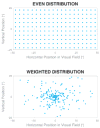
Charts showing simulated phosphene distribution patterns. Vertical and horizontal axes represent the positions along the visual field, and each dot represents a phosphene from an independent electrode contact. While there are equal numbers of contacts in the 2 diagrams, the lower pattern matches the intrinsic acuity profile of the primate visual system, and has a much higher focal acuity than the upper pattern. Upper: When prosthesis electrode contacts are placed on the retina in a regular pattern, the generated phosphenes also appear in a regular pattern across the visual field. Lower: When contacts are placed in a physically regular pattern in tissue downstream of the retina, the phosphenes will appear in a pattern strongly weighted to the center of the visual field.
Fig. 4
Fig. 4
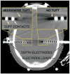
Computed tomography scan showing the implanted depth electrodes. Depth electrodes similar to ones that would be appropriate for use in an LGN-based visual prosthesis are already in clinical use during preparation for surgical treatment of epilepsy such as shown in this image obtained in a patient with bilaterally implanted hippocampus. Left inset: A depth electrode that combines traditional cuff-style contacts with a central bundle of microwires exiting distally. Right inset: A traditional depth electrode without the central bundle.

Similar articles

Cited by

References

    1. Andrews TJ, Halpern SD, Purves SD. Correlated size variations in human visual cortex, lateral geniculate nucleus, and optic tract. J Neurosci. 1997;17:2859–2868. - PMC - PubMed
    1. Bak M, Girvin JP, Hambrecht FT, Kufta CV, Loeb GE, Schmidt EM. Visual sensations produced by intracortical microstimulation of the human occipital cortex. Med Biol Eng Comput. 1990;28:257–259. - PubMed
    1. Bradley DC, Troyk PR, Berg JA, Bak M, Cogan S, Erickson R, et al. Visuotopic mapping through a multichannel stimulating implant in primate V1. J Neurophysiol. 2005;93:1659–1670. - PubMed
    1. Brelén ME, De Potter P, Gersdorff M, Cosnard G, Veraart C, Delbeke J. Intraorbital implantation of a stimulating electrode for an optic nerve visual prosthesis. J Neurosurg. 2006;104:593–597. - PubMed
    1. Brindley GS. Physiology of the Retina and the Visual Pathway. London: Edward Arnold Ltd; 1960.