Comparative Study
. 2009 May 20;1(2):e00009.
doi: 10.1042/AN20090002.

Brain injury expands the numbers of neural stem cells and progenitors in the SVZ by enhancing their responsiveness to EGF

Affiliations
Comparative Study

Brain injury expands the numbers of neural stem cells and progenitors in the SVZ by enhancing their responsiveness to EGF

Dhivyaa Alagappan et al. ASN Neuro. .

Abstract

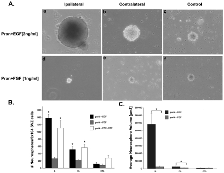
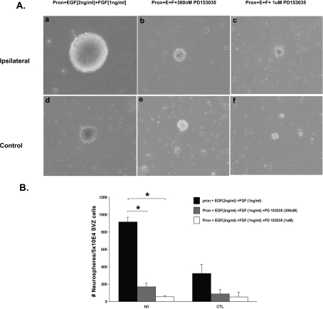
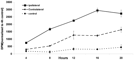
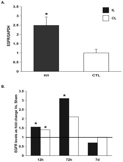
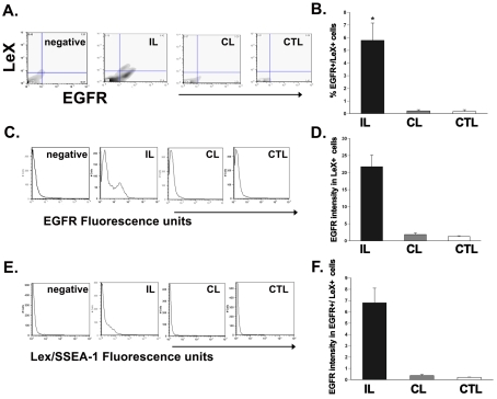
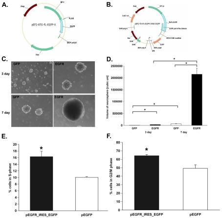
There is an increase in the numbers of neural precursors in the SVZ (subventricular zone) after moderate ischaemic injuries, but the extent of stem cell expansion and the resultant cell regeneration is modest. Therefore our studies have focused on understanding the signals that regulate these processes towards achieving a more robust amplification of the stem/progenitor cell pool. The goal of the present study was to evaluate the role of the EGFR [EGF (epidermal growth factor) receptor] in the regenerative response of the neonatal SVZ to hypoxic/ischaemic injury. We show that injury recruits quiescent cells in the SVZ to proliferate, that they divide more rapidly and that there is increased EGFR expression on both putative stem cells and progenitors. With the amplification of the precursors in the SVZ after injury there is enhanced sensitivity to EGF, but not to FGF (fibroblast growth factor)-2. EGF-dependent SVZ precursor expansion, as measured using the neurosphere assay, is lost when the EGFR is pharmacologically inhibited, and forced expression of a constitutively active EGFR is sufficient to recapitulate the exaggerated proliferation of the neural stem/progenitors that is induced by hypoxic/ischaemic brain injury. Cumulatively, our results reveal that increased EGFR signalling precedes that increase in the abundance of the putative neural stem cells and our studies implicate the EGFR as a key regulator of the expansion of SVZ precursors in response to brain injury. Thus modulating EGFR signalling represents a potential target for therapies to enhance brain repair from endogenous neural precursors following hypoxic/ischaemic and other brain injuries.


Figures

Figure 1
Figure 1. Neurospheres grow larger in the presence of EGF, but not FGF-2
(A) SVZ cells were dissociated from ipsilateral (a and d), contralateral (b and e) and control (c and f) hemispheres at 3 days recovery from H/I and cultured for 7 DIV in ProN medium supplemented with either 2 ng/ml EGF (a–c) or 1 ng/ml FGF-2 (d–f). Phase-contrast images of representative neurospheres were captured for quantification. (B) Quantification of the average number of neurospheres obtained per 50 000 SVZ cells. (C) Quantification of the average neurosphere size from at least 30 spheres. *P<0.05 using ANOVA and Tukey's post-hoc tests. Values are the mean number/size of neurospheres±S.E.M. of three experiments with six animals per experiment. IL, ipsilateral; CL, contralateral; CTL, control.
Figure 2
Figure 2. Pharmacologically inhibiting EGFR abrogates the in vitro NSP response observed following neonatal H/I
(A) SVZ cells were dissociated from ipsilateral (a–c) and control (d–f) hemispheres at 3 days recovery from H/I and cultured for 7 DIV in ProN medium supplemented with 2 ng/ml EGF or 1 ng/ml FGF-2 along with the EGFR inhibitor PD153035 at 300 nM (b and e) or 1 μM (c and f) or with no inhibitor (a and d). (B) Quantification of the average number of neurospheres generated from ipsilateral and control SVZ after 72 h recovery from H/I upon culturing in growth-factor-supplemented medium with or without the EGFR-specific inhibitor PD153035. Values are the means±S.E.M. of three independent experiments with n = 6 animals per experiment. *P<0.05, as measured using a Student's t test.
Figure 3
Figure 3. NSPs proliferate more rapidly in the presence of EGF following neonatal H/I
Animals were killed 3 days after H/I and neurospheres were cultured from ipsilateral (▪), contralateral (♦) and control (•) SVZs in 20 ng/ml EGF overnight. The following morning [3H]thymidine was added to the culture medium (8 μCi/ml). At 4 h intervals, cells were collected on Whatman filter discs by vacuum, the DNA was precipitated with TCA and [3H]thymidine incorporation was quantified. This Figure shows one representative experiment from three repetitions, normalized to the 4 h control. Values are means±S.E.M.
Figure 4
Figure 4. H/I increases EGFR mRNA levels
Ipsilateral H/I and control SVZs were dissected and total RNA was extracted and amplified by (A) semi-quantitative PCR using primers specific for EGFR and normalized to the expression of GAPDH (glyceraldehyde-3-phosphate dehydrogenase) after 72 h of recovery; (B) real-time PCR using primers specific for EGFR and normalized to expression of 18S after 2 h, 72 h and 7 day recovery (n = 6). The solid line represents EGFR expansion in sham-operated animals. *P<0.05 as measured using the Student's t test (A) and REST (B).
Figure 5
Figure 5. H/I increases the levels of activated EGFR protein
(AC) Cryostat sections (12 μm) from animals killed 48 h after H/I by intracardiac perfusion were stained for EGFR and counterstained with DAPI. Panels are representative of the ipsilateral hemisphere (A), contralateral hemisphere (B) and an immunostaining control (C). The scale bar represents 40 μm. To evaluate EGFR activation, H/I and control SVZs were dissected after 72 h recovery and the membranes were subfractionated (see the Materials and methods section) and 30 μg of each membrane fraction was analysed by Western blot for total EGFR and for phosphorylated EGFR (D). The Western blots were quantified and normalized to total receptor levels (E). Results are averaged from three independent experiments (n = 6). *P<0.05 (as measured using a Student's t test).
Figure 6
Figure 6. H/I increases the number of EGFR-positive NG2-expressing neural precursors in the rat SVZ
(A) Dot plot of EGFR+/NG2+ cells from the ipsilateral, contralateral and control SVZs after 72 h recovery from H/I. (B) Quantification of the EGFR+/NG2+ neural precursors in the SVZ. (C) Quantification of EGFR+/NG2 population, encompassing neural stem cells, in the SVZ after injury. Values are the mean number±S.E.M. and are from one representative experiment (n = 6 animals per experiment). *P<0.05, measured using one-way ANOVA and Tukey's post-hoc tests. IL, ipsilateral; CL, contralateral; CTL, control.
Figure 7
Figure 7. H/I increases the number of EGFR+ PSA-NCAM-expressing progenitor neuroblasts in the rat SVZ
(A) Dot plot of EGFR+/PSA-NCAM+ cells from the ipsilateral, contralateral and control SVZs after 72 h recovery from H/I. (B) Quantification of the EGFR+/ PSA-NCAM+ neural precursor population in the SVZ. (C) Quantification of the EGFR+/PSA-NCAM population, encompassing neural stem cells, in the SVZ after injury. Values represent the mean number±S.E.M. and are from one representative experiment with n = 6 animals per experiment. *P<0.05, measured using one-way ANOVA and Tukey's post-hoc tests. IL, ipsilateral; CL, contralateral; CTL, control.
Figure 8
Figure 8. H/I increases the EGFR sensitivity on neural precursors in the rat SVZ
(A) Histogram plot of EGFR intensity within the neural-precursor-positive cells from the ipsilateral, contralateral and control SVZs after 72 h recovery from H/I. (B) Quantification of the EGFR intensity (in a.u.) within the neural precursor population in the SVZ after injury. (C) Quantification of the EGFR intensity (in a.u.) within the neural-precursor-negative, putative stem-cell-encompassing population in the SVZ after injury. Values are the mean number±S.E.M. of three independent experiments with n = 6 animals per experiment. *P<0.05, measured using one-way ANOVA and Tukey's post-hoc tests. IL, ipsilateral; CL, contralateral; CTL, control.
Figure 9
Figure 9. H/I increases the expression of EGFR-positive LeX-expressing putative stem cells in murine SVZ
(A) Dot plot of EGFR+/LeX+ cells from the ipsilateral, contralateral and control SVZs after 72 h recovery from H/I. (B) Quantification of the EGFR+/LeX+ neural stem population in the SVZ after 72 h recovery from H/I. (C) Histogram plot of EGFR intensity within the LeX-positive cells from the ipsilateral, contralateral and control SVZs after 72 h recovery from H/I. (D) Quantification of the EGFR intensity (in a.u.) within the LeX-positive stem cell population in the SVZ after injury. (E) Histogram plot of LeX intensity from the ipsilateral, contralateral and control SVZs after 72 h recovery from H/I. (F) Quantification of the LeX intensity (in a.u.) of SVZ cells after injury. Values represent the mean number±S.E.M. of three independent experiments with n = 6 animals per experiment. *P<0.05, measured using one-way ANOVA and Tukey's post-hoc tests. IL, ipsilateral; CL, contralateral; CTL, control.
Figure 10
Figure 10. EGFR overexpression is sufficient to increase cell-cycle kinetics of SVZ NSPs
(A and B) pEF2-ATG-FL-EGFP and pEF2-fl-Fc-EGFR-IRES-EGFP plasmids. (C) Neurospheres generated at 3 days and 7 days following transfection with pEGFP and pEGFR-IRES-EGFP into SVZ NSPs. (D) Quantification of neurosphere size obtained from (C). (E) Percentage of cells in S-phase after a 6 h BrdU pulse and in G2/M-phase (F). Values represent the mean number±S.E.M. of three independent experiments with n = 6 animals per experiment. *P<0.05, measured using a Student's t test.

References

    1. Aguirre A, Gallo V. Postnatal neurogenesis and gliogenesis in the olfactory bulb from NG2-expressing progenitors of the subventricular zone. J Neurosci. 2004;24:10530–10541. - PMC - PubMed
    1. Aguirre A, Rizvi TA, Ratner N, Gallo V. Overexpression of the epidermal growth factor receptor confers migratory properties to nonmigratory postnatal neural progenitors. J Neurosci. 2005;25:11092–11106. - PMC - PubMed
    1. Alagappan D. Brain injury expands the numbers of neural stem cells and progenitors in the SVZ by enhancing their responsiveness to EGF. Ph.D. Thesis, UMDNJ New Jersey Medical School, Graduate School of Biomedical Sciences. 2008 - PMC - PubMed
    1. Anton ES, Ghashghaei HT, Weber JL, McCann C, Fischer TM, Cheung ID, Gassmann M, Messing A, Klein R, Schwab MH, Lloyd KCK, Lai C. Receptor tyrosine kinase ErbB4 modulates neuroblast migration and placement in the adult forebrain. Nat Neurosci. 2004;7:1319–1328. - PubMed
    1. Arvidsson A, Collin T, Kirik D, Kokaia Z, Lindvall O. Neuronal replacement from endogenous precursors in the adult brain after stroke. Nat Med. 2002;8:963–970. - PubMed

Publication types