

Papillary fibroelastoma of the left atrial wall: a case report
- PMID: 19570232
- PMCID: PMC2715399
- DOI: 10.1186/1749-8090-4-28
Papillary fibroelastoma of the left atrial wall: a case report
Abstract
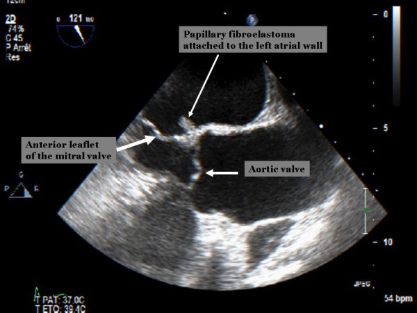
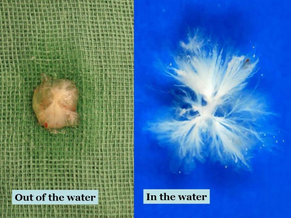
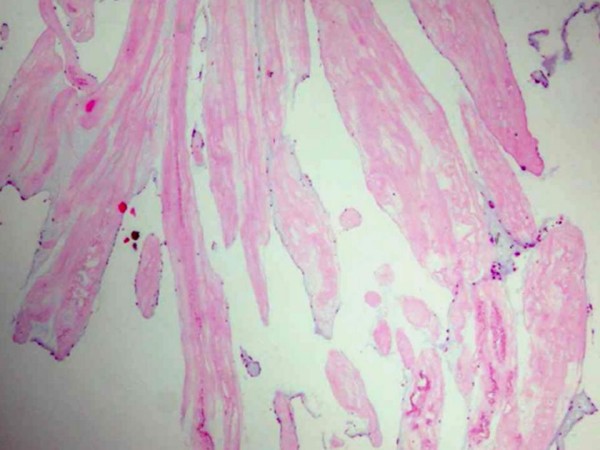
Cardiac papillary fibroelastoma is a rare, benign cardiac tumor. It often arises from valvular endocardium, and non-valvular endocardial location is rare. Although transthoracic echocardiography is usually sufficient for the diagnosis of most cardiac tumors, small tumors such as papillary fibroelastoma may be missed. Transesophageal echocardiography is superior to transthoracic echocardiography in diagnosing these tumors. Despite their benign histology, and independent of their size, they should be resected surgically because of their high potential for embolization. In this report, we present a case of papillary fibroelastoma located on the left atrial wall, presenting with symptoms of cerebral ischemia. The patient was treated surgically for the prevention of further embolic complications. Pertinent literature is also reviewed for this rare and benign cardiac tumor.
Figures
References
-
- Tazelaar HD, Locke TJ, McGregor CGA. Pathology of surgically excised primary cardiac tumors. Mayo Clin Proc. 1992;67:957–65. - PubMed
Publication types
MeSH terms
LinkOut - more resources
Full Text Sources
