

Review
doi: 10.1016/j.stem.2009.06.016.
Control of stem cell fate by physical interactions with the extracellular matrix
Affiliations
- PMID: 19570510
- PMCID: PMC2768283
- DOI: 10.1016/j.stem.2009.06.016
Item in Clipboard
Review
Control of stem cell fate by physical interactions with the extracellular matrix
Cell Stem Cell.
.
Abstract
A diverse array of environmental factors contributes to the overall control of stem cell activity. In particular, new data continue to mount on the influence of the extracellular matrix (ECM) on stem cell fate through physical interactions with cells, such as the control of cell geometry, ECM geometry/topography at the nanoscale, ECM mechanical properties, and the transmission of mechanical or other biophysical factors to the cell. Here, we review some of the physical processes by which cues from the ECM can influence stem cell fate, with particular relevance to the use of stem cells in tissue engineering and regenerative medicine.
Figures
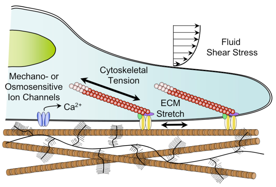
During development and through life, stem cells may be exposed to a variety of physical signals, including tensile, compressive, shear, osmotic, and fluid stresses, often arising secondarily to biomechanical interactions with their ECM. For example, tension of the ECM can induce stretch of the cytoskeleton, and nucleus through focal adhesions, while compression of the ECM can significantly alter local charge density and ion concentrations, potentially activating osmotically sensitive ion channels. Previous studies have shown that these mechanical stimuli individually can strongly influence stem cell growth and differentiation in vivo and in vitro.
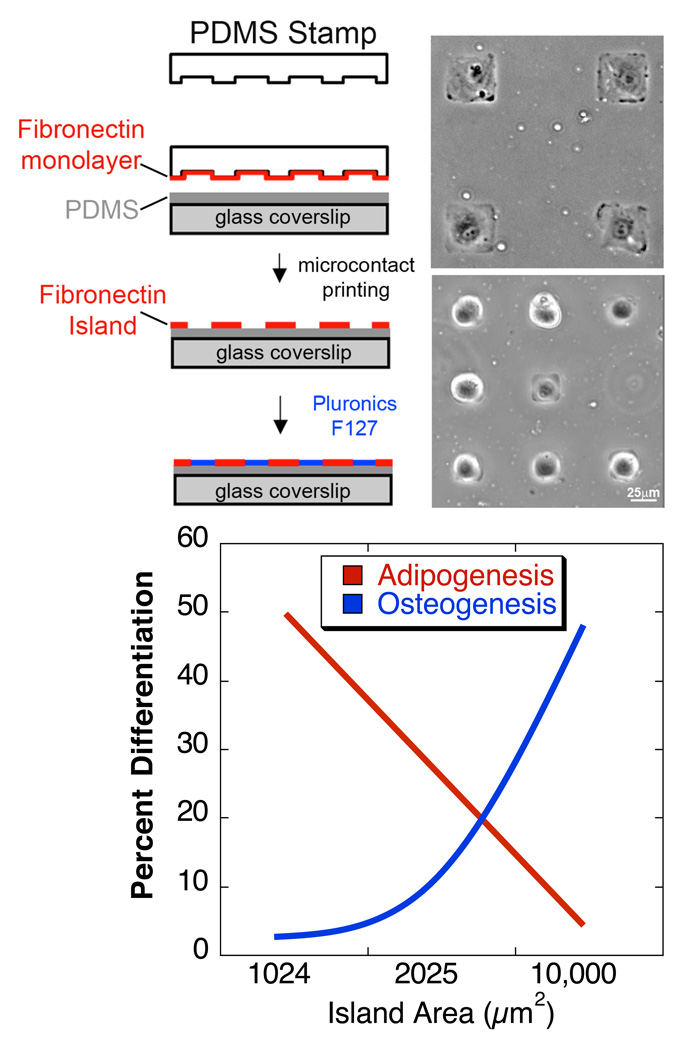
(top) Polydimethylsiloxane (PDMS) stamps with micron-sized features are coated with fibronectin, or other ECM proteins. Fibronectin is transferred from the raised features on the stamp to a PDMS-coated glass coverslip substrate via microcontact printing. Gaps between fibronectin islands are passivated to prevent cell adhesion by adsorption of the non-adhesive, Pluronic F127. By controlling the size of the islands where cells can attach, their shape can be predefined. Phase images (courtesy of R. Desai) of cells patterned on 50×50 or 25×25 µm2 islands. (bottom) human MSCs that were allowed to adhere, flatten, and spread underwent osteogenesis, while unspread, round cells underwent adipogenesis. This switch in lineage commitment was regulated by cell shape through the modulation of endogenous RhoA activity [Data from (McBeath et al., 2004)].
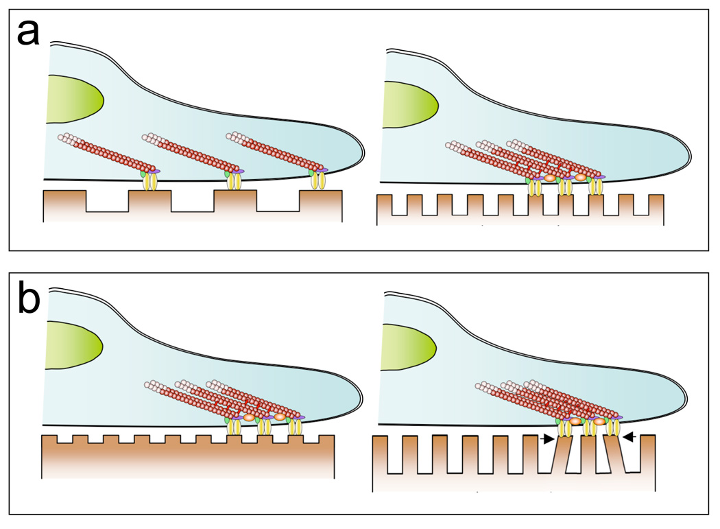
The nanoscale geometry and size of the features of the ECM may have significant effects on a number of cell properties, such as attachment/adhesion, migration, and proliferation, although the mechanisms responsible for these effects are not well understood. (a) Whereas changes in the feature size of the substrate on the scale of single cells could impact adhesion by altering the degree of cell spreading (Figure 2), varying features at the scale of individual adhesions may alter the clustering of integrins and other cell adhesion molecules. Altered clustering can influence the number and distribution of focal adhesions and subsequently, the structure of the cytoskeleton (Arnold et al., 2004). In turn, these factors may further influence cytoskeletal tension and the transmission and transduction of other molecular and biomechanical signals. (b) Differences in the size and structure (e.g., height) of nanotopographic features may influence cell behavior through secondary effects, such as alterations in the effective stiffness of the substrate (e.g. (Discher et al., 2005; Saha et al., 2008a)).
References
-
- Adams GB, Scadden DT. A niche opportunity for stem cell therapeutics. Gene Ther. 2008;15:96–99. - PubMed
-
- Alhadlaq A, Mao JJ. Mesenchymal stem cells: isolation and therapeutics. Stem Cells Dev. 2004;13:436–448. - PubMed
-
- Angele P, Yoo JU, Smith C, Mansour J, Jepsen KJ, Nerlich M, Johnstone B. Cyclic hydrostatic pressure enhances the chondrogenic phenotype of human mesenchymal progenitor cells differentiated in vitro. J Orthop Res. 2003;21:451–457. - PubMed
-
- Arnold M, Cavalcanti-Adam EA, Glass R, Blummel J, Eck W, Kantlehner M, Kessler H, Spatz JP. Activation of integrin function by nanopatterned adhesive interfaces. Chemphyschem. 2004;5:383–388. - PubMed
Publication types
MeSH terms
Grants and funding
- AR048852/AR/NIAMS NIH HHS/United States
- AG15768/AG/NIA NIH HHS/United States
- R01 AR048852/AR/NIAMS NIH HHS/United States
- P41 EB001046/EB/NIBIB NIH HHS/United States
- R01 AR048182/AR/NIAMS NIH HHS/United States
- R01 EB000262/EB/NIBIB NIH HHS/United States
- EB00262/EB/NIBIB NIH HHS/United States
- GM74048/GM/NIGMS NIH HHS/United States
- EB001046/EB/NIBIB NIH HHS/United States
- R01 GM074048/GM/NIGMS NIH HHS/United States
- AR50245/AR/NIAMS NIH HHS/United States
- P01 AR050245/AR/NIAMS NIH HHS/United States
- R01 AG015768/AG/NIA NIH HHS/United States
- AR48182/AR/NIAMS NIH HHS/United States
LinkOut - more resources
Full Text Sources
Other Literature Sources
Medical
