. 2009 Jul 2;5(1):64-75.
doi: 10.1016/j.stem.2009.04.003.

TAp63 prevents premature aging by promoting adult stem cell maintenance

Affiliations

TAp63 prevents premature aging by promoting adult stem cell maintenance

Xiaohua Su et al. Cell Stem Cell. .

Abstract

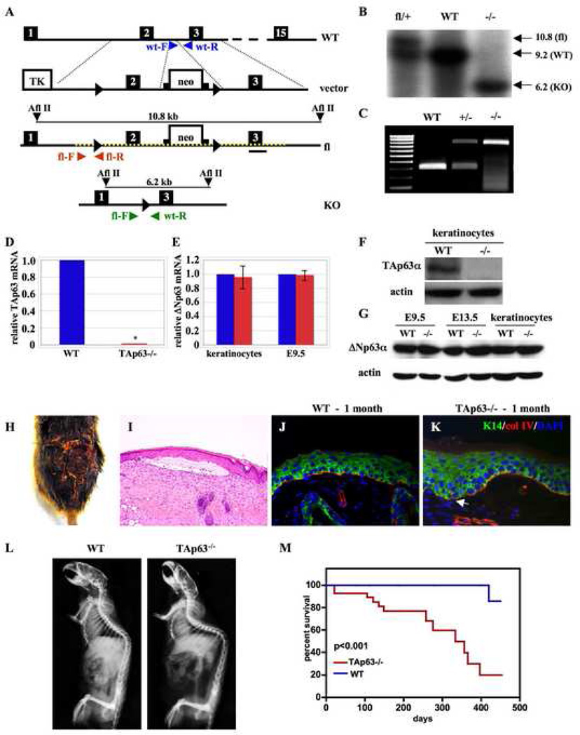
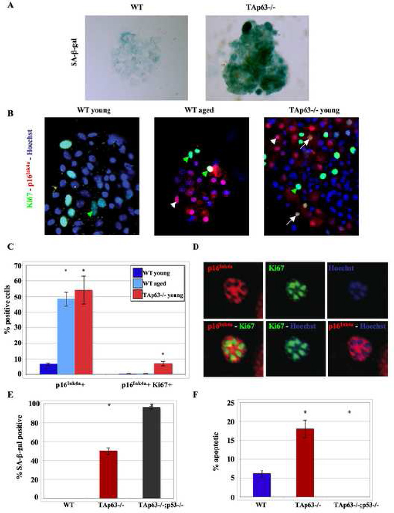
The cellular mechanisms that regulate the maintenance of adult tissue stem cells are still largely unknown. We show here that the p53 family member, TAp63, is essential for maintenance of epidermal and dermal precursors and that, in its absence, these precursors senesce and skin ages prematurely. Specifically, we have developed a TAp63 conditional knockout mouse and used it to ablate TAp63 in the germline (TAp63(-/-)) or in K14-expressing cells in the basal layer of the epidermis (TAp63(fl/fl);K14cre+). TAp63(-/-) mice age prematurely and develop blisters, skin ulcerations, senescence of hair follicle-associated dermal and epidermal cells, and decreased hair morphogenesis. These phenotypes are likely due to loss of TAp63 in dermal and epidermal precursors since both cell types show defective proliferation, early senescence, and genomic instability. These data indicate that TAp63 serves to maintain adult skin stem cells by regulating cellular senescence and genomic stability, thereby preventing premature tissue aging.


Figures

Figure 1
Figure 1. TAp63−/− mice develop blisters and exhibit accelerated aging phenotypes
(A) Generation of the TAp63 conditional knockout mice. The TAp63 targeting vector was generated by inserting loxP sites (triangles) flanking exon 2 and a neomycin (neo) cassette flanked by frt sites (squares) in intron 2. Primers used for genotyping by PCR of wild-type (blue) and TAp63 conditional knockout (fl) (red) alleles are shown. The targeted region of the floxed allele is depicted by a dashed yellow line. (B) Southern blot analysis of genomic DNA from TAp63fl/+, WT, and TAp63−/− mice. (C) PCR of DNA from wild-type (WT), TAp63+/− (+/−) and TAp63−/− (−/−) mice. (D, E) Quantitative RT-PCR of (D) TAp63 mRNA from wild-type (WT) and TAp63−/− keratinocytes or of (E) ΔNp63 mRNA from wild-type (blue) and TAp63−/− (red) keratinocytes and day 9.5 (E9.5) embryos. GAPDH was used as an internal control. (F, G) Western blot for TAp63 (F) in wild-type (WT) and TAp63−/− passage 2 keratinocytes or for ΔNp63 (G) in wild-type (WT) and TAp63−/− E9.5 and E13.5 embryos and keratinocytes. Actin was used as an internal control. (H) Dorsum of an 8-month TAp63−/− mouse with an ulcerated wound. (I) H&E cross section of skin from a 1 month TAp63−/− mouse with a blister. Arrows indicate the split at the epidermal-dermal junction with entrapped serum. (J, K) Immunocytochemistry for keratin 14 (green) and collagen IV (red) of skin from 1 month wild-type (J) and TAp63−/− mice (K). DAPI was used as a counterstain. Magnification 200X. White arrow indicates the beginning of the dermal/epidermal separation. N=6 mice per group. (L) X-ray of 8 month wild-type (WT) and TAp63−/− mice. (M) Kaplan-Meier survival curve for wild-type (WT) and TAp63−/− mice. Median survival for TAp63−/− and WT mice is 333 and 712 days, respectively, n=29 mice per group, p<0.001.
Figure 2
Figure 2. TAp63−/− mice lose hair follicles with age
(A) Confocal images of immunocytochemistry of a hair follicle from a young, wild-type mouse for fibronectin (green), NCAM (dark blue), p63 (red), and Hoechst 33258 (blue). Hatched line denotes the dermal papilla (DP), yellow arrowhead indicates a p63-positive dermal sheath (DS) cell, arrows show a p63-positive dermal papilla cells, and white arrowheads epidermal cells with nuclear p63. Magnification 630X. (B) RT-PCR for TAp63 mRNA in Sox2:EGFP-positive (+ve) and negative (−ve) cells from adult backskin. #1, 2 and 3 are samples from 3 different mice, and the ungated sample is total skin cells from one mouse. (C) SA-β-gal staining (blue) of hair follicles in young wild-type or TAp63−/− skin (left two panels). The arrows indicate SA-β-gal-positive dermal papillae. The right two panels, (a) and (b), are micrographs of sequential sections through the same hair follicle, 40µm apart. The arrowhead indicates an SA-β-gal-positive dermal sheath cell (DS), and the arrows a positive dermal papilla (DP). (D) Quantification of SA-β-gal positive cells in the dermal papillae (DP). n = 3 animals per group. *p<0.05. (E) H&E stained skin sections of 1, 6, and 12 month wild-type (WT) and TAp63−/− mice. Arrows denote hair follicles, and double-arrows indicate regions of TAp63−/− skin depleted of hair follicles. Magnification 100X. (F) Quantification of hair follicles per millimeter (E). N = 8 and 4 mice each for wild-type (WT) and TAp63−/− at 1 month and 12 months, respectively. *p<0.05. (G) Quantification of hair follicles in wild-type (WT) and TAp63−/− mice at 1, 6–9, and 12 months. Only patches of skin with no follicles for greater than 400µm were included. N = 4 mice of each genotype at each time point. *p<0.05.
Figure 3
Figure 3. TAp63−/− mice have an impaired wound-healing response
(A,B) Photomicrographs of wild-type (A) and TAp63−/− (B) H&E stained skin sections 6 days after wounding. Dotted lines indicate the dermal-epidermal boundary and boxes denote area of immunohistochemical analysis on serial sections shown at higher magnification in panels D, E, G, H, I, & J. (C) Bar graph indicating relative wound size 6 days after wounding in wild-type (WT), TAp63−/− (−/−), TAp63fl/fl;K14cre− (K14cre−), and TAp63fl/fl;K14cre+ (K14cre+) mice. N=6 mice per group, *p<0.05. (D,E) Immunohistochemistry for BrdU incorporation 6 days after wounding in wild-type (D) and TAp63−/− (E) mice. (F) Quantification of BrdU incorporation in epidermal cells in sections shown in panels D,E in WT, TAp63−/−, TAp63fl/fl;K14cre−, and TAp63fl/fl;K14Cre+ mice. N=6 mice per group, *p<0.001. (G–J) Immunohistochemistry for keratin 5 (G,H; brown) or TAp63 (I,J; brown) in wild-type (G,I) or TAp63−/− (H,J) skin. (K) Quantification of BrdU incorporation in dermal cells. In panels (D, E, G, H, I, J), tissue was counterstained with hematoxylin. Arrowheads denote BrdU positive dermal and epidermal cells (D, E) and TAp63-positive epidermal cells (I). N=6 mice per genotype. (L) qRT-PCR of mRNA from WT, TAp63−/−, and TAp63fl/fl;K14cre+ epidermal cells extracted from E18.5 skin. N=6 mice per genotype in triplicate. (M) Western blots for TAp63 and p53 in stressed keratinocytes (passage 2) and freshly-isolated keratinocytes (passage 0) from wild-type (WT) and TAp63−/− mice. (N) Western blot for TAp63 in WT, TAp63−/−, and TAp63fl/fl;K14cre+ stressed keratinocytes (passage 2). Actin was used as an internal control. Keratinocytes derived from 3 independent embryos for each genotype and performed in triplicate.
Figure 4
Figure 4. TAp63 regulates colony formation from epidermal precursors
(A) Epidermal colonies from wild-type (WT), TAp63fl/fl, TAp63−/−, TAp63−/−;p53−/− and TAp63fl/fl;K14Cre+ mice cultured for 8 or 12 days on J2 3T3 feeder layers and stained with rhodamine B. (B) Immunostaining for BrdU (green) and K5 (red) in wild-type and TAp63−/− epidermal clones. (C,D) Quantification of BrdU incorporation in colonies after 8 (C) or 20 (D) days in culture. *p<0.05. N = 3 cell lines per genotype in triplicate. (E) Immunostaining for the spinous marker, K10 (green), in wild-type (WT) and TAp63−/− colonies cultured in high calcium (1.2 mM) media for 2 days. DAPI was used as a counterstain.
Figure 5
Figure 5. TAp63 regulates self-renewal of dermal precursors via p57Kip2
(A, B) RT-PCR in WT and p63−/− SKPs for (A) TAp63 and ΔNp63 mRNAs and (B) p53, p73, and GAPDH mRNAs. Embryonic day 18 cortical tissue (C) was used as a positive control. (C) Immunocytochemistry for p63 (green) in SKP spheres isolated from WT skin, from reconstituted hair follicles (WT follicle), or from E18 p63−/− skin. Hoechst (blue) used as a counterstain. Magnification 400X. (D) SKP spheres isolated from young WT or TAp63−/− skin, or from E18 p63−/− skin and immunostained for fibronectin, nestin, vimentin, smooth muscle actin (SMA) and versican. Magnification 400X. (E) SKP spheres isolated from neonatal WT skin or from E18 p63−/− skin, immunostained for Ki67 (red), and counterstained with Hoechst (blue). Magnification 400X. (F) Quantification of Ki67 positive cells. n = 3, *p < 0.05. (G) Quantification of cells that form a new sphere at clonal density (2500 cells/ml) in methylcellulose cultures. n = 3, *p < 0.05. (H) RT-PCR analysis for p57Kip2 and GAPDH mRNAs in E18 cortex (C), young WT and TAp63−/− SKPs and E18 p63−/− SKPs. (I) SKP spheres isolated from young WT and TAp63−/− skin, immunostained for p57Kip2 (red) and counterstained for Hoechst (blue). Magnification 400X. (J) Quantification of p57Kip2 positive cells. n = 3, *p < 0.05. (K) ChIP for p63 in young WT or E18 p63−/− SKPs and PCR for the p57Kip2 core promoter. (L) Immunostaining for p57Kip2 (red) and Ki67 (green) in TAp63−/− SKPs transfected with or without a p57Kip2 expression vector. Hoechst (blue) was used as a counterstain. Transfected p57Kip2-positive cells were negative for Ki67. Magnification 200X, n=3.
Figure 6
Figure 6. Loss of TAp63 causes dermal precursor cells to senesce
(A) SKP spheres from young WT and TAp63−/− SKPs stained for SA-β-gal (blue). Magnification 400X. (B) SKP spheres from young and aged WT and young TAp63−/− skin immunostained for p16Ink4a (red) and Ki67 (green) and counterstained with Hoechst (blue). Green arrowheads indicate Ki67, white arrowheads indicate p16Ink4a, and white arrows indicate Ki67 and p16Ink4a double positive cells. Magnification 400X. (C) Quantification of p16Ink4a-positive and p16Ink4a, Ki67 double-positive cells. n = 3, *p < 0.05. (D) High magnification of a TAp63−/− SKP cell nucleus that was labeled for p16Ink4a (red) and Ki67 (green) and counterstained with Hoechst (blue). (E,F) Quantification of SA-β-gal positive (E) or apoptotic (F) keratinocytes in wild-type (WT), TAp63−/−, and TAp63−/−;p53−/− cultures. N=3 cell lines for each genotype, performed in triplicate. *p<0.0001.
Figure 7
Figure 7. Loss of TAp63 leads to genomic instability
(A) SKP spheres from young and aged WT and TAp63−/− skin, immunostained for γ-H2AX (green) and counterstained for Hoechst (blue). Magnification 400X. (B) Quantification of γ-H2AX positive cells. n = 3, *p < 0.05. (C) Immunostaining for γ-H2AX (green) in aged wild-type and young TAp63−/− skin and counterstained Hoechst (blue). Arrows denote hair follicles, and arrowheads indicate positive cells within the dermal sheath. Magnification 200X. (D) Immunostaining for γ-H2AX foci (red) in wild-type and TAp63−/− keratinocytes. DAPI was used as a counterstain. (E) Metaphase spreads of wild-type and TAp63−/− primary keratinocytes (passage 0). Cytogenetic aberrations in TAp63−/− cells are indicated by colored arrows. Chromosomal fusions (black arrows), fragments (red arrows), breaks (green arrowheads), biarmed chromosomes (blue arrow), ring chromosome (blue arrowhead).

Comment in

References

    1. Ackerman AB. Histologic Diagnosis of Inflammatory Skin Diseases. 2nd Ed. Baltimore, MD: Williams & Wilkins; 1997.
    1. Barrandon Y, Green H. Three clonal types of keratinocyte with different capacities for multiplication. Proceedings of the National Academy of Sciences of the United States of America. 1987;84:2302–2306. - PMC - PubMed
    1. Beretta C, Chiarelli A, Testoni B, Mantovani R, Guerrini L. Regulation of the cyclin-dependent kinase inhibitor p57Kip2 expression by p63. Cell Cycle. 2005;4:1625–1631. - PubMed
    1. Biernaskie JA, McKenzie IA, Toma JG, Miller FD. Isolation of skin-derived precursors (SKPs) and differentiation and enrichment of their Schwann cell progeny. Nat Protoc. 2006;1:2803–2812. - PubMed
    1. Candi E, Rufini A, Terrinoni A, Dinsdale D, Ranalli M, Paradisi A, De Laurenzi V, Spagnoli LG, Catani MV, Ramadan S, et al. Differential roles of p63 isoforms in epidermal development: selective genetic complementation in p63 null mice. Cell Death and Differentiation. 2006;13:1037–1047. - PubMed

Publication types

MeSH terms