

Arterial gene transfer of the TGF-beta signalling protein Smad3 induces adaptive remodelling following angioplasty: a role for CTGF
- PMID: 19570811
- PMCID: PMC2761202
- DOI: 10.1093/cvr/cvp220
Arterial gene transfer of the TGF-beta signalling protein Smad3 induces adaptive remodelling following angioplasty: a role for CTGF
Abstract
Aims: Although transforming growth factor-beta (TGF-beta) is believed to stimulate intimal hyperplasia after arterial injury, its role in remodelling remains unclear. We investigate whether Smad3, a TGF-beta signalling protein, might facilitate its effect on remodelling.
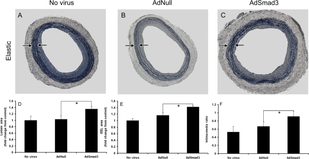
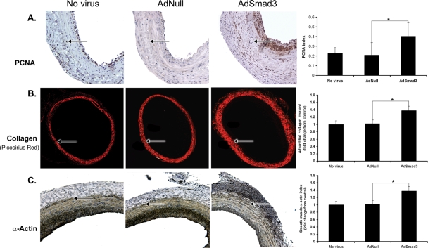
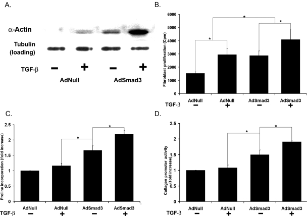
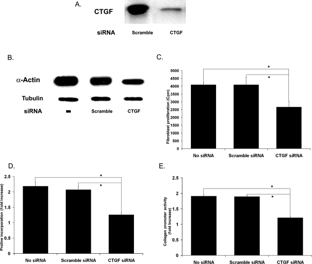
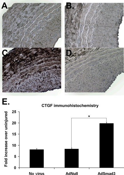
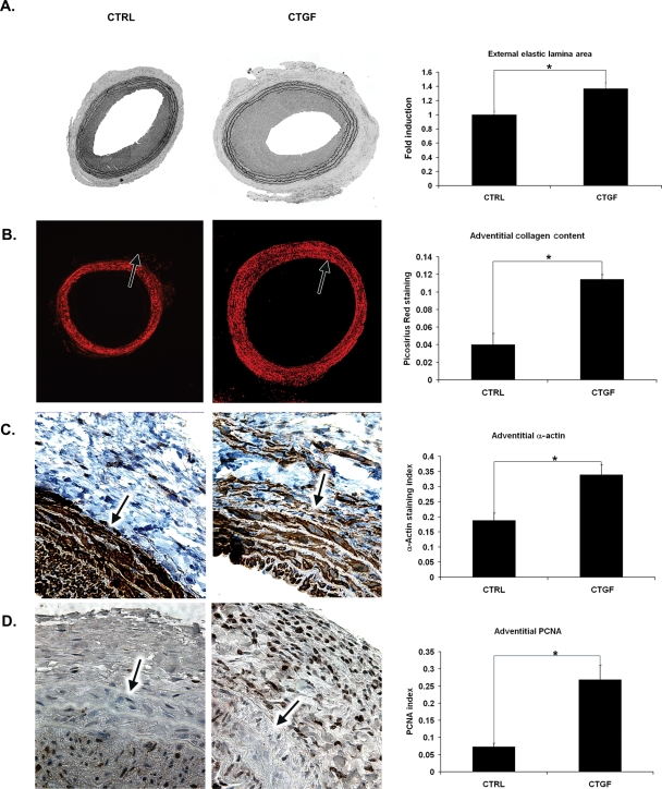
Methods and results: Using the rat carotid angioplasty model, we assess Smad3 expression following arterial injury. We then test the effect of arterial Smad3 overexpression on the response to injury, and use a conditioned media experimental design to confirm an Smad3-dependent soluble factor that mediates this response. We use small interfering RNA (siRNA) to identify this factor as connective tissue growth factor (CTGF). Finally, we attempt to replicate the effect of medial Smad3 overexpression through adventitial application of recombinant CTGF. Injury induced medial expression of Smad3; overexpression of Smad3 caused neointimal thickening and luminal expansion, suggesting adaptive remodelling. Smad3 overexpression, though exclusively medial, caused adventitial changes: myofibroblast transformation, proliferation, and collagen production, all of which are associated with adaptive remodelling. Supporting the hypothesis that Smad3 initiated remodelling and these adventitial changes via a secreted product of medial smooth muscle cells (SMCs), we found that media conditioned by Smad3-expressing recombinant adenoviral vector (AdSmad3)-infected SMCs stimulated adventitial fibroblast transformation, proliferation, and collagen production in vitro. This effect was attenuated by pre-treatment of SMCs with siRNA specific for CTGF, abundantly produced by AdSmad3-infected SMCs, and significantly up-regulated in Smad3-overexpressing arteries. Moreover, periadventitial administration of CTGF replicated the effect of medial Smad3 overexpression on adaptive remodelling and neointimal hyperplasia.
Conclusion: Medial gene transfer of Smad3 promotes adaptive remodelling by indirectly influencing the behaviour of adventitial fibroblasts. This arterial cell-cell communication is likely to be mediated by Smad3-dependent production of CTGF.
Figures
Similar articles
-
Preferential secretion of collagen type 3 versus type 1 from adventitial fibroblasts stimulated by TGF-β/Smad3-treated medial smooth muscle cells.Cell Signal. 2013 Apr;25(4):955-60. doi: 10.1016/j.cellsig.2012.12.021. Epub 2012 Dec 29. Cell Signal. 2013. PMID: 23280188 Free PMC article.
-
TGF-beta through Smad3 signaling stimulates vascular smooth muscle cell proliferation and neointimal formation.Am J Physiol Heart Circ Physiol. 2009 Aug;297(2):H540-9. doi: 10.1152/ajpheart.91478.2007. Epub 2009 Jun 12. Am J Physiol Heart Circ Physiol. 2009. PMID: 19525370 Free PMC article.
-
Protein kinase C-delta mediates adventitial cell migration through regulation of monocyte chemoattractant protein-1 expression in a rat angioplasty model.Arterioscler Thromb Vasc Biol. 2012 Apr;32(4):943-54. doi: 10.1161/ATVBAHA.111.244921. Epub 2012 Feb 9. Arterioscler Thromb Vasc Biol. 2012. PMID: 22328773 Free PMC article.
-
NADPH oxidase 4 contributes to connective tissue growth factor expression through Smad3-dependent signaling pathway.Free Radic Biol Med. 2016 May;94:174-84. doi: 10.1016/j.freeradbiomed.2016.02.031. Epub 2016 Mar 3. Free Radic Biol Med. 2016. PMID: 26945889
-
Heterogeneous subpopulations of adventitial progenitor cells regulate vascular homeostasis and pathological vascular remodelling.Cardiovasc Res. 2022 May 6;118(6):1452-1465. doi: 10.1093/cvr/cvab174. Cardiovasc Res. 2022. PMID: 33989378 Free PMC article. Review.
Cited by
-
Aortic response to balloon injury in obese Zucker rats.Comp Med. 2012 Aug;62(4):264-70. Comp Med. 2012. PMID: 23043778 Free PMC article.
-
TGF-β/Smad3 stimulates stem cell/developmental gene expression and vascular smooth muscle cell de-differentiation.PLoS One. 2014 Apr 9;9(4):e93995. doi: 10.1371/journal.pone.0093995. eCollection 2014. PLoS One. 2014. PMID: 24718260 Free PMC article.
-
AAV2/8-hSMAD3 gene delivery attenuates aortic atherogenesis, enhances Th2 response without fibrosis, in LDLR-KO mice on high cholesterol diet.J Transl Med. 2014 Sep 20;12:252. doi: 10.1186/s12967-014-0252-8. J Transl Med. 2014. PMID: 25236373 Free PMC article.
-
Halofuginone stimulates adaptive remodeling and preserves re-endothelialization in balloon-injured rat carotid arteries.Circ Cardiovasc Interv. 2014 Aug;7(4):594-601. doi: 10.1161/CIRCINTERVENTIONS.113.001181. Epub 2014 Jul 29. Circ Cardiovasc Interv. 2014. PMID: 25074254 Free PMC article.
-
Local CXCR4 Upregulation in the Injured Arterial Wall Contributes to Intimal Hyperplasia.Stem Cells. 2016 Nov;34(11):2744-2757. doi: 10.1002/stem.2442. Epub 2016 Jul 17. Stem Cells. 2016. PMID: 27340942 Free PMC article.
References
-
- Post MJ, Borst C, Kuntz RE. The relative importance of arterial remodeling compared with intimal hyperplasia in lumen renarrowing after balloon angioplasty. A study in the normal rabbit and the hypercholesterolemic Yucatan micropig. Circulation. 1994;89:2816–2821. - PubMed
-
- Ward MR, Pasterkamp G, Yeung AC, Borst C. Arterial remodeling. Mechanisms and clinical implications. Circulation. 2000;102:1186–1191. - PubMed
-
- Kakuta T, Currier JW, Haudenschild CC, Ryan TJ, Faxon DP. Differences in compensatory vessel enlargement, not intimal formation, account for restenosis after angioplasty in the hypercholesterolemic rabbit model. Circulation. 1994;89:2809–2815. - PubMed
-
- Losordo DW, Rosenfield K, Kaufman J, Pieczek A, Isner JM. Focal compensatory enlargement of human arteries in response to progressive atherosclerosis. In vivo documentation using intravascular ultrasound. Circulation. 1994;89:2570–2577. - PubMed
-
- Nakamura Y, Zhao H, Yutani C, Imakita M, Ishibashi-Ueda H. Morphometric and histologic assessment of remodeling associated with restenosis after percutaneous transluminal coronary angioplasty. Cardiology. 1998;90:115–121. - PubMed
Publication types
MeSH terms
Substances
Grants and funding
LinkOut - more resources
Full Text Sources
Other Literature Sources
Medical
Molecular Biology Databases
Miscellaneous
