

doi: 10.1128/JVI.01127-09.
Epub 2009 Jul 1.
Prion infectivity in fat of deer with chronic wasting disease
Affiliations
- PMID: 19570855
- PMCID: PMC2738259
- DOI: 10.1128/JVI.01127-09
Item in Clipboard
Prion infectivity in fat of deer with chronic wasting disease
J Virol.
2009 Sep.
Abstract
Chronic wasting disease (CWD) is a neurodegenerative prion disease of cervids. Some animal prion diseases, such as bovine spongiform encephalopathy, can infect humans; however, human susceptibility to CWD is unknown. In ruminants, prion infectivity is found in central nervous system and lymphoid tissues, with smaller amounts in intestine and muscle. In mice, prion infectivity was recently detected in fat. Since ruminant fat is consumed by humans and fed to animals, we determined infectivity titers in fat from two CWD-infected deer. Deer fat devoid of muscle contained low levels of CWD infectivity and might be a risk factor for prion infection of other species.
Figures
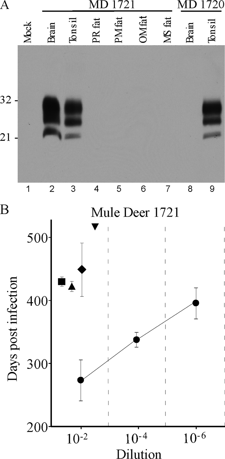
(A) Western blot of PrPres from CWD agent-infected mule deer. All samples were treated with proteinase K as described in the text. Lanes 1 and 4 to 8 were loaded with 2-mg tissue equivalents. Lanes 2, 3, and 9 were loaded with 0.5-mg tissue equivalents. Lane 1 contains uninfected deer brain, lanes 2 to 7 show tissues from mule deer (MD) 1721, and lanes 8 and 9 show tissues from mule deer 1720. The blot was probed using the anti-PrP antibody 6H4 (Prionics) by enhanced-chemiluminescence detection. Film exposure was 14 min. Numbers at the left are molecular weights (in thousands). PR, perirenal; PM, perimuscular; OM, omental; MS, mediastinal. (B) CWD incubation periods in TgDeerPrP mice following intracerebral injection of tissue homogenates from mule deer 1721. Symbols: •, brain; ▪, perirenal fat; ▴, perimuscular fat; ♦, omental fat; ▾, mediastinal fat (only one of four mice developed CWD, so there are no error bars). Note that the incubation periods found in fat at a 10−2 dilution are slightly longer than the incubation periods following inoculation of a 10−6 dilution of brain, suggesting 10,000- to 100,000-fold-lower infectivity in fat than in brain.
References
-
- Angers, R. C., S. R. Browning, T. S. Seward, C. J. Sigurdson, M. W. Miller, E. A. Hoover, and G. C. Telling. 2006. Prions in skeletal muscles of deer with chronic wasting disease. Science 311:1117. - PubMed
-
- Beringue, V., J. L. Vilotte, and H. Laude. 2008. Prion agent diversity and species barrier. Vet. Res. 39:47. - PubMed
-
- Bradley, R. 1996. Bovine spongiform encephalopathy distribution and update on some transmission and decontamination studies, p. 11-28. In C. J. Gibbs, Jr. (ed.), Bovine spongiform encephalopathy. The BSE dilemma. Springer-Verlag, New York, NY.
-
- Buschmann, A., and M. H. Groschup. 2005. Highly bovine spongiform encephalopathy-sensitive transgenic mice confirm the essential restriction of infectivity to the nervous system in clinically diseased cattle. J. Infect. Dis. 192:934-942. - PubMed
-
- Chesebro, B., M. Trifilo, R. Race, K. Meade-White, C. Teng, R. Lacasse, L. Raymond, C. Favara, G. Baron, S. Priola, B. Caughey, E. Masliah, and M. Oldstone. 2005. Anchorless prion protein results in infectious amyloid disease without clinical scrapie. Science 308:1435-1439. - PubMed
Publication types
MeSH terms
Grants and funding
LinkOut - more resources
Full Text Sources
