. 2009 Sep;83(18):9163-74.
doi: 10.1128/JVI.01979-08. Epub 2009 Jul 1.

Distinctive effects of the Epstein-Barr virus family of repeats on viral latent gene promoter activity and B-lymphocyte transformation

Affiliations

Distinctive effects of the Epstein-Barr virus family of repeats on viral latent gene promoter activity and B-lymphocyte transformation

Ahmed K M Ali et al. J Virol. 2009 Sep.

Abstract

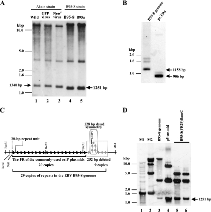
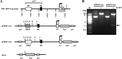
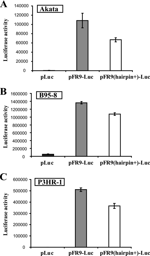
The Epstein-Barr virus (EBV), a human B-lymphotropic gamma herpesvirus, contains multiple repetitive sequences within its genome. A group of repetitive sequences, known as the family of repeats (FR), contains multiple binding sites for the viral trans-acting protein EBNA-1. The FR sequences are important for viral genome maintenance and for the regulation of the promoter involved in viral latent gene expression. It has been reported that a palindromic sequence with a putative secondary structure exists at the 3' end of the FR in the genome of the EBV B95-8 strain and that this palindromic sequence has been deleted from the FR of the commonly used EBV miniplasmids. For the first time, we cloned an EBV B95-8 DNA fragment containing the full-length FR, which enabled us to examine the functional difference between full-length and deleted FRs. The full-length FR, like the deleted FR, functioned as a transcriptional enhancer of the viral latent gene promoter, but that transactivation was significantly attenuated in the case of the full-length FR. No significant enhancement of replication was observed when the deleted FR was replaced with the full-length FR in an EBV miniplasmid. By contrast, when the same set of FR sequences were tested in the context of the complete EBV genome, the full-length FR resulted in more-efficient B-cell transformation than the deleted FR. We propose that the presence of the full-length FR contributes to the precise regulation of the viral latent promoter and increases the efficiency of B-cell transformation.


Figures

FIG. 1.
FIG. 1.
(A) Southern blot analysis demonstrating the stability of the sizes of the FRs of the different EBV strains in latently infected cells. The results of the Akata cells harboring the wild-type Akata EBV (Wild, lane 1), two Akata derivatives harboring either a recombinant EBV expressing GFP (24) (GFP virus, lane 2) or a recombinant EBV expressing the neomycin resistance gene (39) (Neor virus, lane 3), B95-8 cells (lane 4), and B95a cells (lane 5) are shown. The calculated sizes of the FR fragments (EcoRI-MluI fragments) are also indicated. (B) The sizes of the FRs in the EBV B95-8 strain genome and in pCEP4 were examined by Southern blot analysis (XcmI-MluI digestion) by using the XcmI-MluI fragment of pCEP4 as a probe. The calculated sizes of the XcmI-MluI fragments (containing the FR) are indicated. (C) Schematic representation of the full-length FR and the deleted FR. Each arrow represents a 30-bp repeat unit. The region of the 252-bp sequence (nine copies of repeats with a 128-bp palindromic sequence) that is missing from the 3′ end of the FR of the commonly used oriP plasmids is indicated. The restriction endonuclease sites of EcoRI, XcmI, NsiI, BstXI, EcoRV, and MluI are indicated. Note that 30-bp repeat units around the palindromic sequence are inversely oriented (5). (D) Successful cloning of the full-length FR of EBV B95-8 strain into a cloning vector. The Southern blot results of the B95-8 genomic DNA (lane 3), the p5 cosmid (lane 4), and B95-8(FR29)-BamC (lanes 5 and 6) are shown. Signals hybridizing to DNA size markers (M1 and M2) (lanes 1 and 2) and the calculated size of the full-length FR fragments are indicated.
FIG. 2.
FIG. 2.
(A) Schematic diagrams of the reporter constructs containing either the deleted or the full-length FR. The locations of oriP and the Cp promoter region of EBV B95-8 strain DNA are indicated on top. Two functional elements of the oriP (the FR and DS), the transcription start site of Cp (arrows), the luciferase reporter gene (Luc), and polyadenylation signals (pA) are indicated. C1 and C2 (top) are exons giving rise to the leader sequence of EBNA-encoding messages. pFR20-Luc harbors the FR with 20 copies of 30-bp repeats, while pFR29-Luc harbors the FR with 29 copies of 30-bp repeats (FR29, open box). pLuc serves as a control plasmid having only the SfiI synthetic linker (white arrowhead) instead of oriP and Cp. Restriction endonuclease sites are shown for BstXI (B), EcoRV (E), NsiI (N), and SfiI (Sf). (B) Plasmid DNAs of pFR20-Luc or pFR29-Luc, either undigested (−) or digested with EcoRV (+), were electrophoresed along with a DNA size marker (M) in 0.8% agarose gel. The EcoRV fragment (1,122 bp) being excised from pFR29-Luc is indicated by an arrow.
FIG. 3.
FIG. 3.
The enhancer activities of either the deleted FR (FR20) or the full-length FR (FR29) were determined by luciferase reporter assay. EBV-positive Akata cells (A), B95-8 cells (B), P3HR-1 cells (C), and BJAB cells (D) were used as recipient cells for transfection. Indicated amounts of an EBNA-1 expression plasmid were cotransfected together with the reporter constructs in the case of panel D. The results are expressed as the mean values ± the standard errors of the means (n = 5 in panels A and B and 3 in panels C and D).
FIG. 4.
FIG. 4.
(A) Schematic diagrams of the reporter constructs having nine copies of 30-bp repeats with and without a putative secondary structure. Plasmids pFR20-Luc and pFR29-Luc are identical to those depicted in Fig. 2A, and the DS of oriP, the transcription start site of Cp, the first exon (C1), the luciferase gene (Luc), and polyadenylation signals (pA) are also as depicted in Fig. 2A. pFR9-Luc harbors the first nine copies of FR that are not expected to form any secondary structure, while pFR9(hairpin+)-Luc contains the last nine copies of the full-length FR that possibly forms a stable stem-loop structure (5). The 273-bp sequences that have been artificially deleted during the plasmid construction are indicated. Restriction endonuclease sites are shown for BstXI (B), EcoRV (E), NsiI (N), and SpeI (Sp). (B, C) Plasmid DNAs of pFR9-Luc and pFR9(hairpin+)-Luc, either undigested (−) or enzyme-digested (+) (NsiI-SpeI [B] and EcoRV [C]), were electrophoresed along with a DNA size marker (M) in 0.8% agarose gel. The NsiI-SpeI fragments being excised from both of the plasmids are indicated by an arrowhead in panel B, while the EcoRV fragment being excised only from pFR9(hairpin+)-Luc is indicated by an arrow in panel C.
FIG. 5.
FIG. 5.
The reporter construct having nine copies of the 30-bp repeat, which possibly forms a secondary structure, exhibits attenuated transactivation in EBV latently infected B cells. EBV-positive Akata cells (A), B95-8 cells (B), and P3HR-1 cells (C) were used as recipient cells for the luciferase reporter assay. The results are expressed as the mean values ± standard errors of the means (n = 4 for panel A and 5 for panels B and C).
FIG. 6.
FIG. 6.
Replication activities of the test plasmids after being transiently introduced into Raji cells. Hirt extracts of transduced Raji cells were digested with restriction enzymes as indicated, and the digested DNA samples were analyzed by Southern blotting. The DNA fragment of an ampicillin resistance gene was used as a probe. The results obtained by SspI-DpnI digestion (top blot) or SspI digestion alone (bottom blot) are shown. The test plasmids, either with the deleted FR (pFR20-ΔLuc) or the full-length FR (pFR29-ΔLuc), and a control plasmid (pΔLuc) are indicated at the top. Two samples per test plasmid were analyzed, as the experiment was performed in a duplicated manner. In the top blot, only the top bands representing the DpnI-resistant replicated molecules are shown.
FIG. 7.
FIG. 7.
(A) Verification of the FR sizes of the test BACmids. DNA samples were simultaneously digested by NsiI and MluI (indicated in Fig. 1C) and analyzed by Southern hybridization by using the FR20 sequence as a probe. The calculated sizes of the NsiI-MluI fragments are indicated. (B) DNA restriction enzyme mapping of the test BACmids. DNAs of AK-BAC-GFP, FR20-BAC (two independent clones), and FR29-BAC (two independent clones) were digested by either BamHI or NcoI, and the digested samples were analyzed by 0.8% agarose gel electrophoresis. The bands containing the FR region are indicated by white dots. The bands representing the IR1 are also indicated. (C) Comparable GFP-inducing titers of virus mixtures containing either FR20-BAC virus or FR29-BAC virus. EBV-negative Daudi cells were infected with serially diluted (10-fold or 100-fold) mixture viruses containing either FR20-BAC or FR29-BAC. Infected cells were analyzed by fluorescence-activated cell sorting using FL1 and FL2 channels, and GFP-expressing cells were identified by the shift of fluorescence intensity in the FL1 channel. Numbers represent the percentages of GFP-positive cells (surrounded by dotted line) after infection. The result of uninfected cells is also shown (top left). (D) PCR amplification of DNA from the EBNA-3C coding region of the latently infected EBV genomes in the established LCLs. The PCR products of the Akata EBV genome (type 1 EBV) and those of the P3HR-1 EBV genome (type 2 EBV) served as controls. (E) Five independent LCLs harboring only type 1 EBV were selected for FR20-BAC and FR29-BAC, respectively, and BAC clones were rescued from such LCLs. Three independent BAC clones from each LCL were digested by BamHI and analyzed. The BamHI C fragments containing the FR region are indicated by white dots, while the bands representing terminal repeats are indicated by black dots. Note the size difference between the BamHI C fragments of FR20-BAC (indicated by a black arrow) and those of FR29-BAC (comigrating with BamHI B fragment, indicated by a white arrowhead).

Similar articles

Cited by

References

    1. Ambinder, R. F., W. A. Shah, D. R. Rawlins, G. S. Hayward, and S. D. Hayward. 1990. Definition of the sequence requirements for binding of the EBNA-1 protein to its palindromic target sites in Epstein-Barr virus DNA. J. Virol. 64:2369-2379. - PMC - PubMed
    1. Baer, R., A. T. Bankier, M. D. Biggin, P. L. Deininger, P. J. Farrell, T. J. Gibson, G. Hatfull, G. S. Hudson, S. C. Satchwell, C. Seguin, et al. 1984. DNA sequence and expression of the B95-8 Epstein-Barr virus genome. Nature 310:207-211. - PubMed
    1. Bodescot, M., M. Perricaudet, and P. J. Farrell. 1987. A promoter for the highly spliced EBNA family of RNAs of Epstein-Barr virus. J. Virol. 61:3424-3430. - PMC - PubMed
    1. Floettmann, J. E., K. Ward, A. B. Rickinson, and M. Rowe. 1996. Cytostatic effect of Epstein-Barr virus latent membrane protein-1 analyzed using tetracycline-regulated expression in B cell lines. Virology 223:29-40. - PubMed
    1. Fruscalzo, A., G. Marsili, V. Busiello, L. Bertolini, and D. Frezza. 2001. DNA sequence heterogeneity within the Epstein-Barr virus family of repeats in the latent origin of replication. Gene 265:165-173. - PubMed

Publication types

MeSH terms

Substances

LinkOut - more resources