. 2009 Sep;83(18):9247-57.
doi: 10.1128/JVI.00840-09. Epub 2009 Jul 1.

A simian immunodeficiency virus-infected macaque model to study viral reservoirs that persist during highly active antiretroviral therapy

Affiliations

A simian immunodeficiency virus-infected macaque model to study viral reservoirs that persist during highly active antiretroviral therapy

Jason B Dinoso et al. J Virol. 2009 Sep.

Abstract

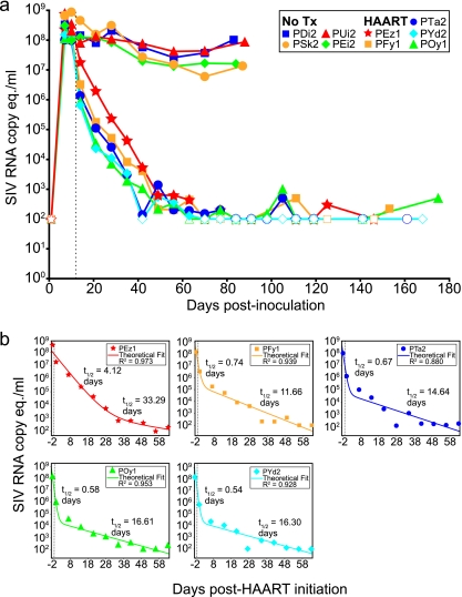
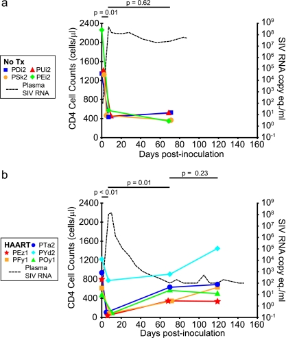
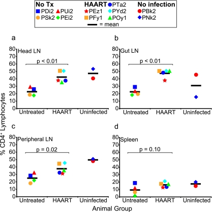
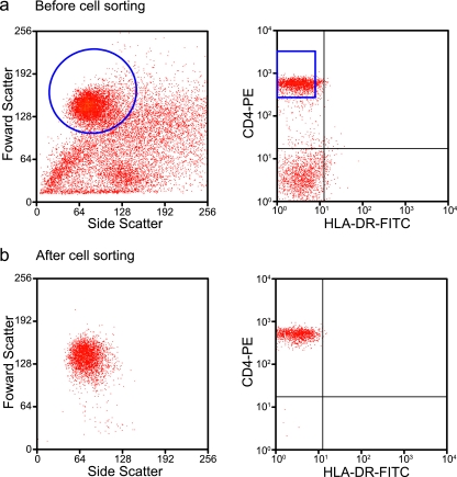
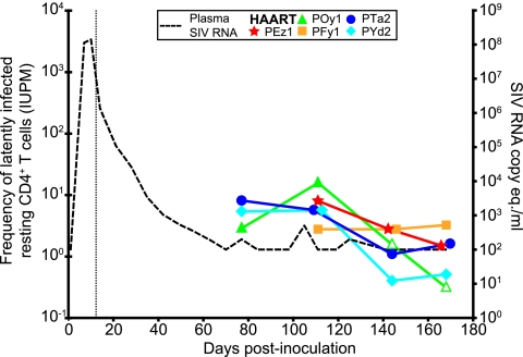
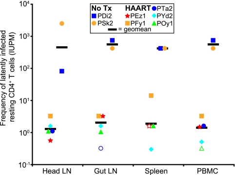
The treatment of human immunodeficiency virus type 1 (HIV-1) infection with highly active antiretroviral therapy (HAART), a combination of three or more antiretroviral drugs, suppresses viremia below the clinical limit of detection (50 HIV-1 RNA copies/ml), but latently infected resting CD4(+) T cells serve as lifelong reservoirs, and low-level viremia can be detected with special assays. Recent studies have provided evidence for additional reservoirs that contribute to residual viremia but are not present in circulating cells. Identification of all the sources of residual viremia in humans may be difficult. These discoveries highlight the need for a tractable model system to identify additional viral reservoirs that could represent barriers to eradication. In this study, simian immunodeficiency virus (SIV)-infected pig-tailed macaques (Macaca nemestrina) were treated with four antiretroviral drugs to develop an animal model for viral suppression during effective HAART. Treatment led to a biphasic decay in viremia and a significant rise in levels of circulating CD4(+) T cells. At terminal infection time points, the frequency of circulating resting CD4(+) T cells harboring replication-competent virus was reduced to a low steady-state level similar to that observed for HIV-infected patients on HAART. The frequencies of resting CD4(+) T cells harboring replication-competent virus in the pooled head lymph nodes, gut lymph nodes, spleen, and peripheral blood were reduced relative to those for untreated SIV-infected animals. These observations closely parallel findings for HIV-infected humans on suppressive HAART and demonstrate the value of this animal model to identify and characterize viral reservoirs persisting in the setting of suppressive antiretroviral drugs.


Figures

FIG. 1.
FIG. 1.
Suppression of SIV viremia with HAART. (a) Levels of viremia were measured from day 0 to the day of necropsy for untreated animals (No Tx) (n = 4) and HAART-treated animals (n = 5). In the HAART-treated animals, viremia was reduced at least by ∼5 log10 units relative to the untreated animals, to levels below the limit of detection. Uninfected animals (n = 2) were consistently negative for SIV RNA. (b) Mathematical modeling of the decay of viremia for each animal revealed a biphasic decay that occurred upon initiation of HAART. Half-lives (t1/2) for each phase of decay are shown for each animal. These values are similar to half-lives reported for humans initiating HAART. Open symbols represent measurements at or below the limit of detection (100 SIV RNA copy equivalents [eq.]/ml). The vertical dotted line represents the day when HAART was initiated (day 12) for the treated animals.
FIG. 2.
FIG. 2.
Effect of HAART on CD4+ T-cell counts in peripheral blood. Counts of circulating CD4+ T cells were measured at days 0, 7, and 70 p.i. for both untreated (No Tx) (n = 4) (a) and HAART-treated (n = 5) (b) animals and at day 119 for HAART-treated animals. For both untreated and HAART-treated animals, cell counts were significantly reduced between day 0 and day 7, the time interval during which viremia peaks. CD4+ T-cell counts were significantly increased by day 70 in the HAART-treated animals but not in untreated animals. Changes in cell counts between day 70 and day 119 were not significant.
FIG. 3.
FIG. 3.
Fraction of CD4+ T cells in tissues. The percentage of lymphocytes that were CD4+ was measured in head lymph nodes (LN) (cervical, retropharyngeal, and submandibular) (a), gut lymph nodes (mesenteric and colonic) (b), peripheral lymph nodes (axillary and inguinal) (c), and spleen (d) in untreated (n = 4), HAART-treated (n = 5), and uninfected (n = 2) animals. Differences between untreated (No Tx) and HAART-treated animals were significant for the lymph node tissue but not spleen. Mean measurements are indicated by horizontal bars.
FIG. 4.
FIG. 4.
Isolation of resting CD4+ T cells. Resting CD4+ T cells were isolated from blood and various lymphoid tissues by magnetic bead depletion followed by sorting for small morphologically homogeneous populations of CD4+ HLA-DR cells. (a) After magnetic bead enrichment, enriched cells contained a morphologically heterogeneous population of cells (left) and some CD4 cells (right). (b) Upon cell sorting using CD4-PE and HLA-DR-fluorescein isothiocyanate (FITC), isolated cells were morphologically (left) and phenotypically (CD4+ HLA-DR) (right) homogeneous, with 98 to 99% purity.
FIG. 5.
FIG. 5.
Frequency of circulating resting CD4+ T cells harboring replication-competent virus. At various times after undetectable viremia (dotted line, right y axis) was achieved with HAART, circulating resting CD4+ T cells (small CD4+ HLA-DR cells) were isolated and assayed for the presence of inducible, replication-competent SIV. For HAART-treated animals, the frequencies of cells harboring replication-competent virus in circulating resting CD4+ T cells isolated at days 77, 111, and 141 and at necropsy (days 161 to 175) were measured. The vertical dotted line represents the day when HAART was initiated (day 12). Open symbols represent measurements less than the indicated values. The confidence intervals at each data point are ±0.7 logs.
FIG. 6.
FIG. 6.
Frequency of resting CD4+ T cells harboring replication-competent virus in tissues and blood at necropsy. Resting CD4+ T cells (small CD4+ HLA-DR cells) were isolated from head lymph nodes (LN), gut lymph nodes, spleen, and PBMC at necropsy. Purified cells were assayed for the presence of cells harboring replication-competent virus. Geometric mean frequencies for HAART-treated animals were ≥2 log10 values lower than those for untreated (No Tx) animals and were not statistically different between tissues (P = 0.47). Open symbols represent measurements less than the indicated values. The confidence intervals at each data point are ±0.7 logs.

References

    1. Ambrose, Z., S. Palmer, V. F. Boltz, M. Kearney, K. Larsen, P. Polacino, L. Flanary, K. Oswald, M. Piatak, Jr., J. Smedley, W. Shao, N. Bischofberger, F. Maldarelli, J. T. Kimata, J. W. Mellors, S. L. Hu, J. M. Coffin, J. D. Lifson, and V. N. KewalRamani. 2007. Suppression of viremia and evolution of human immunodeficiency virus type 1 drug resistance in a macaque model for antiretroviral therapy. J. Virol. 81:12145-12155. - PMC - PubMed
    1. Babas, T., J. B. Dewitt, J. L. Mankowski, P. M. Tarwater, J. E. Clements, and M. C. Zink. 2006. Progressive selection for neurovirulent genotypes in the brain of SIV-infected macaques. AIDS 20:197-205. - PubMed
    1. Babas, T., E. Vieler, D. A. Hauer, R. J. Adams, P. M. Tarwater, K. Fox, J. E. Clements, and M. C. Zink. 2001. Pathogenesis of SIV pneumonia: selective replication of viral genotypes in the lung. Virology 287:371-381. - PubMed
    1. Bailey, J. R., A. R. Sedaghat, T. Kieffer, T. Brennan, P. K. Lee, M. Wind-Rotolo, C. M. Haggerty, A. R. Kamireddi, Y. Liu, J. Lee, D. Persaud, J. E. Gallant, J. Cofrancesco, Jr., T. C. Quinn, C. O. Wilke, S. C. Ray, J. D. Siliciano, R. E. Nettles, and R. F. Siliciano. 2006. Residual human immunodeficiency virus type 1 viremia in some patients on antiretroviral therapy is dominated by a small number of invariant clones rarely found in circulating CD4+ T cells. J. Virol. 80:6441-6457. - PMC - PubMed
    1. Barouch, D. H., J. Kunstman, M. J. Kuroda, J. E. Schmitz, S. Santra, F. W. Peyerl, G. R. Krivulka, K. Beaudry, M. A. Lifton, D. A. Gorgone, D. C. Montefiori, M. G. Lewis, S. M. Wolinsky, and N. L. Letvin. 2002. Eventual AIDS vaccine failure in a rhesus monkey by viral escape from cytotoxic T lymphocytes. Nature 415:335-339. - PubMed

Publication types

MeSH terms