

Anterograde spread of herpes simplex virus type 1 requires glycoprotein E and glycoprotein I but not Us9
- PMID: 19570876
- PMCID: PMC2738194
- DOI: 10.1128/JVI.00633-09
Anterograde spread of herpes simplex virus type 1 requires glycoprotein E and glycoprotein I but not Us9
Abstract
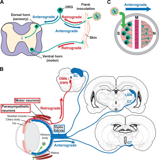
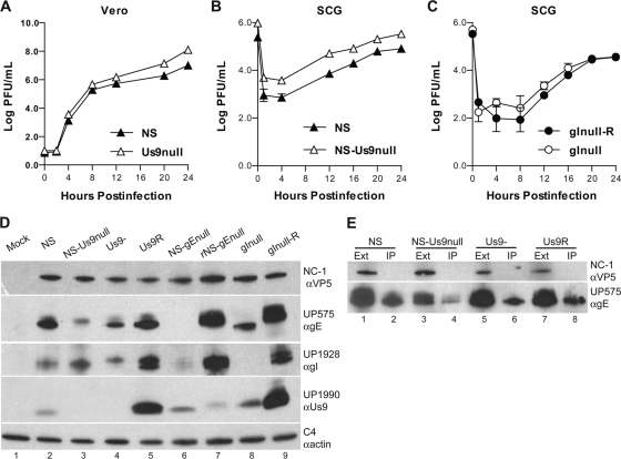
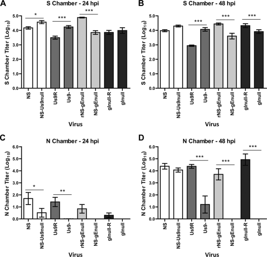
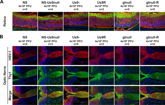
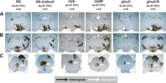
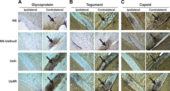
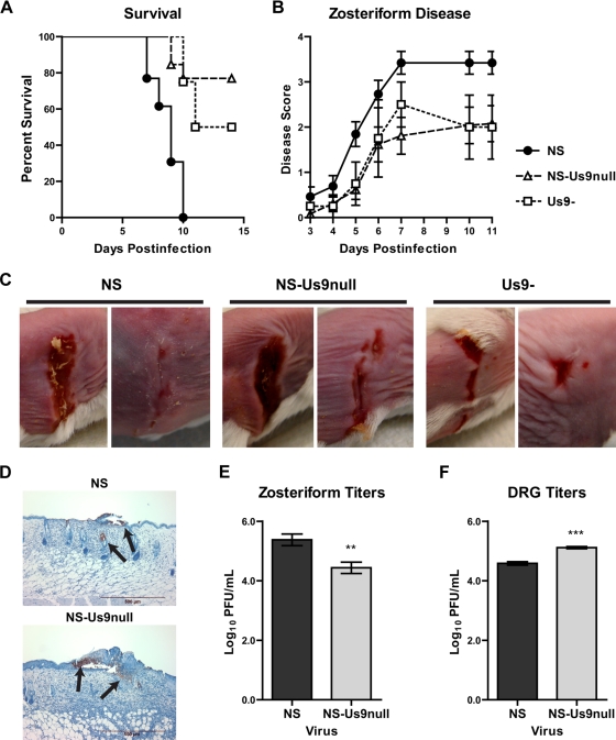
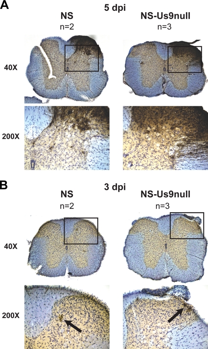
Anterograde neuronal spread (i.e., spread from the neuron cell body toward the axon terminus) is a critical component of the alphaherpesvirus life cycle. Three viral proteins, gE, gI, and Us9, have been implicated in alphaherpesvirus anterograde spread in several animal models and neuron culture systems. We sought to better define the roles of gE, gI, and Us9 in herpes simplex virus type 1 (HSV-1) anterograde spread using a compartmentalized primary neuron culture system. We found that no anterograde spread occurred in the absence of gE or gI, indicating that these proteins are essential for HSV-1 anterograde spread. However, we did detect anterograde spread in the absence of Us9 using two independent Us9-deleted viruses. We confirmed the Us9 finding in different murine models of neuronal spread. We examined viral transport into the optic nerve and spread to the brain after retinal infection; the production of zosteriform disease after flank inoculation; and viral spread to the spinal cord after flank inoculation. In all models, anterograde spread occurred in the absence of Us9, although in some cases at reduced levels. This finding contrasts with gE- and gI-deleted viruses, which displayed no anterograde spread in any animal model. Thus, gE and gI are essential for HSV-1 anterograde spread, while Us9 is dispensable.
Figures
Similar articles
-
Glycoproteins gE and gI are required for efficient KIF1A-dependent anterograde axonal transport of alphaherpesvirus particles in neurons.J Virol. 2013 Sep;87(17):9431-40. doi: 10.1128/JVI.01317-13. Epub 2013 Jun 26. J Virol. 2013. PMID: 23804637 Free PMC article.
-
Herpes Simplex Virus gE/gI and US9 Promote both Envelopment and Sorting of Virus Particles in the Cytoplasm of Neurons, Two Processes That Precede Anterograde Transport in Axons.J Virol. 2017 May 12;91(11):e00050-17. doi: 10.1128/JVI.00050-17. Print 2017 Jun 1. J Virol. 2017. PMID: 28331094 Free PMC article.
-
Herpes simplex virus gE/gI and US9 proteins promote transport of both capsids and virion glycoproteins in neuronal axons.J Virol. 2008 Nov;82(21):10613-24. doi: 10.1128/JVI.01241-08. Epub 2008 Aug 27. J Virol. 2008. PMID: 18753205 Free PMC article.
-
Anterograde transport of α-herpesviruses in neuronal axons.Virology. 2021 Jul;559:65-73. doi: 10.1016/j.virol.2021.02.011. Epub 2021 Mar 4. Virology. 2021. PMID: 33836340 Review.
-
Anterograde Neuronal Circuit Tracers Derived from Herpes Simplex Virus 1: Development, Application, and Perspectives.Int J Mol Sci. 2020 Aug 18;21(16):5937. doi: 10.3390/ijms21165937. Int J Mol Sci. 2020. PMID: 32824837 Free PMC article. Review.
Cited by
-
Investigating the biology of alpha herpesviruses with MS-based proteomics.Proteomics. 2015 Jun;15(12):1943-56. doi: 10.1002/pmic.201400604. Epub 2015 May 15. Proteomics. 2015. PMID: 25764121 Free PMC article. Review.
-
Herpes simplex virus type 2 glycoprotein E is required for efficient virus spread from epithelial cells to neurons and for targeting viral proteins from the neuron cell body into axons.Virology. 2010 Sep 30;405(2):269-79. doi: 10.1016/j.virol.2010.06.006. Epub 2010 Jul 3. Virology. 2010. PMID: 20598729 Free PMC article.
-
Herpes simplex virus 1 pUL34 plays a critical role in cell-to-cell spread of virus in addition to its role in virus replication.J Virol. 2011 Jul;85(14):7203-15. doi: 10.1128/JVI.00262-11. Epub 2011 May 11. J Virol. 2011. PMID: 21561917 Free PMC article.
-
The pseudorabies virus protein, pUL56, enhances virus dissemination and virulence but is dispensable for axonal transport.Virology. 2016 Jan 15;488:179-86. doi: 10.1016/j.virol.2015.11.014. Epub 2015 Dec 1. Virology. 2016. PMID: 26655235 Free PMC article.
-
Glycoproteins gE and gI are required for efficient KIF1A-dependent anterograde axonal transport of alphaherpesvirus particles in neurons.J Virol. 2013 Sep;87(17):9431-40. doi: 10.1128/JVI.01317-13. Epub 2013 Jun 26. J Virol. 2013. PMID: 23804637 Free PMC article.
References
-
- Al-Mubarak, A., and S. I. Chowdhury. 2004. In the absence of glycoprotein I (gI), gE determines bovine herpesvirus type 5 neuroinvasiveness and neurovirulence. J. Neurovirol. 10233-243. - PubMed
-
- Babic, N., B. Klupp, A. Brack, T. C. Mettenleiter, G. Ugolini, and A. Flamand. 1996. Deletion of glycoprotein gE reduces the propagation of pseudorabies virus in the nervous system of mice after intranasal inoculation. Virology 219279-284. - PubMed
-
- Balan, P., N. Davis-Poynter, S. Bell, H. Atkinson, H. Browne, and T. Minson. 1994. An analysis of the in vitro and in vivo phenotypes of mutants of herpes simplex virus type 1 lacking glycoproteins gG, gE, gI or the putative gJ. J. Gen. Virol. 751245-1258. - PubMed
Publication types
MeSH terms
Substances
Grants and funding
LinkOut - more resources
Full Text Sources
