. 2009 Sep;297(3):F662-70.
doi: 10.1152/ajprenal.00146.2009. Epub 2009 Jul 1.

20-HETE activates the Raf/MEK/ERK pathway in renal epithelial cells through an EGFR- and c-Src-dependent mechanism

Affiliations

20-HETE activates the Raf/MEK/ERK pathway in renal epithelial cells through an EGFR- and c-Src-dependent mechanism

Talha Akbulut et al. Am J Physiol Renal Physiol. 2009 Sep.

Abstract

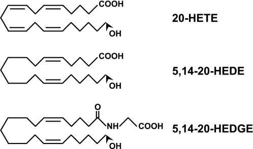
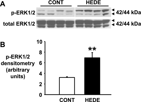
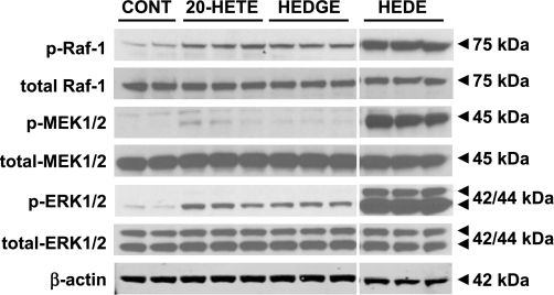
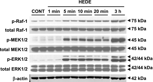
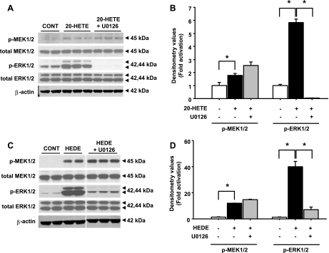
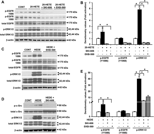
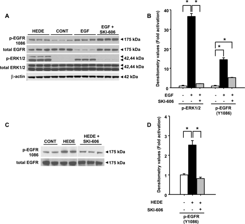
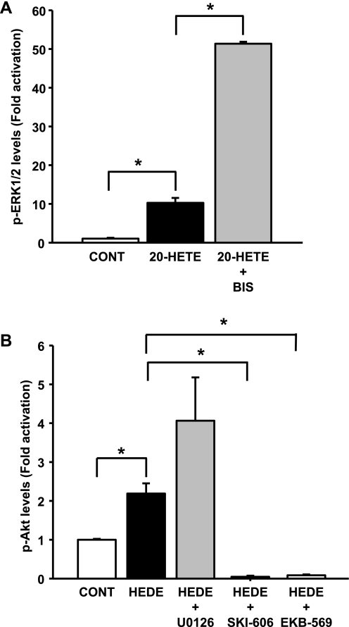
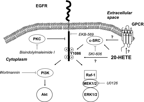
20-Hydroxyeicosatetraenoic acid (20-HETE) has been reported to promote mitogenicity in a variety of cell types, including renal epithelial cells. However, the signal transduction pathways activated by 20-HETE have not been fully defined. The present study evaluated the effects of 20-HETE and its more stable agonist analogs 20-hydroxyeicosa-5(Z),14(Z)-dienoic acid (5,14-20-HEDE) and N-[20-hydroxyeicosa-5(Z),14(Z)-dienoyl]glycine (5,14-20-HEDGE) on the Raf/MEK/ERK and phosphatidylinositol 3-kinase (PI3K)-Akt pathway in LLC-PK(1) renal epithelial cells. 20-HETE (20 microM) increased phosphorylation of Raf-1 (2.5 +/- 0.2-fold), MEK1/2 (6.3 +/- 1.6-fold), and ERK1/2 (5.8 +/- 0.3-fold) compared with vehicle-treated cells. Similarly, the 20-HETE analogs also strongly activated ERK1/2 in a Raf-1- and MEK1/2-dependent manner. Moreover, 5,14-20-HEDE increased Akt phosphorylation by 2.2 +/- 0.3-fold. 20-HETE and 5,14-20-HEDE also promoted activation (Y1086) of epidermal growth factor receptor (EGFR; Y1086) by 1.9 +/- 0.2- and 2.5 +/- 0.2-fold, respectively. These effects were completely blocked by the EGFR inhibitor EKB-569 (0.1 microM). Moreover, EKB-569 (0.1 microM), as well as a c-Src inhibitor, SKI-606 (0.05 microM), completely abolished the 20-HETE-mediated activation of the Raf/MEK/ERK and PI3K-Akt pathways. Blockade of PKC with bisindolylmaleimide I had no effect on 20-HETE-induced ERK1/2 activation. This study demonstrated that 20-HETE activated the Raf/MEK/ERK and Akt pathways in renal epithelial cells secondary to the activation of c-Src and EGFR.


Figures

Fig. 1.
Fig. 1.
Chemical structure of 20-hydroxyeicosatetraenoic acid (20-HETE) and stable 20-HETE mimetics 20-hydroxyeicosa-5(Z),14(Z)-dienoic acid (5,14-20-HEDE) and N-[20-hydroxyeicosa-5(Z),14(Z)-dienoyl]glycine (5,14-20-HEDGE).
Fig. 2.
Fig. 2.
Activation of ERK1/2 in renal medulla of Sprague-Dawley rats after 5,14-20-HEDE treatment. A: representative Western blot depicting ERK1/2 activation after 5,14-20-HEDE (HEDE) treatment (30 mg/kg sc). p-ERK1/2, phosphorylated ERK1/2; CONT, control. B: p-ERK1/2 densitometry (n = 4 individual animals). **Significant difference from control (P < 0.01).
Fig. 3.
Fig. 3.
Activation of Raf/MEK/ERK pathway by 20-HETE and stable 20-HETE mimetics. Representative Western blot demonstrates activation of Raf/MEK/ERK pathway using specific antibodies. Cell lysates were obtained from LLC-PK1 cells incubated in serum-free media containing 20 μM 20-HETE, 20 μM 5,14-20-HEDGE (HEDGE), and 20 μM 5,14-20-HEDE for 3 h. Indomethacin (2 μM) was added to prevent COX-mediated metabolism 60 min before 20-HETE and agonist incubation. Changes in levels of p-ERK1/2, phosphorylated MEK1/2 (p-MEK1/2), and phosphorylated Raf-1 (p-Raf-1) in the presence of 20-HETE, 5,14-20-HEDGE, and 5,14-20-HEDE are shown. β-Actin was used as a loading control. Immunoblots represent 2–8 results for each antibody group.
Fig. 4.
Fig. 4.
Temporal activation of Raf/MEK/ERK pathway by 5,14-20-HEDE. Western blot analysis was performed to determine early activation of Raf/MEK/ERK pathway after incubation of LLC-PK1 cells with 20 μM 5,14-20-HEDE. Cells were collected 1 min, 5 min, 10 min, 20 min, and 3 h after addition of the drug. Cells were treated with vehicle for 5 min to use as control for comparison with 5,14-20-HEDE-treated cells. Blot was performed twice with samples from 2 different batches of cells studied in triplicate for each time point. β-Actin was used as a loading control.
Fig. 5.
Fig. 5.
Effect of MEK inhibition on ERK1/2 activation mediated by 20-HETE and 5,14-20-HEDE. Western blot analysis was performed to determine changes in levels of phosphorylated and total forms of MEK1/2 and ERK1/2 after 3 h of incubation with 20 μM 20-HETE (A and B) and 20 μM 5,14-20-HEDE (C and D) in the presence or absence of 10 μM U0126. B and D: densitometry values for 20-HETE and 5,14-20-HEDE. All bands for each respective antibody were run on the same gel. Bands are separated to indicate that the blot was cropped to bring the most relevant grouping of bands together. Blots were performed twice with the lysates from 2 different batches of cells studied in triplicate for each treatment group. β-Actin was used as a loading control in A and C. *Significant difference between groups (P < 0.05).
Fig. 6.
Fig. 6.
Effect of c-Src and epidermal growth factor receptor (EGFR) inhibition on ERK1/2 activation mediated by 20-HETE and 5,14-20-HEDE. Western blot analysis was performed to determine changes in levels of phosphorylated and total forms of EGFR and ERK1/2 after 3-h incubation with 20 μM 20-HETE (A), EGFR and ERK1/2 after 3-h incubation with 20 μM 5,14-20-HEDE (C), and c-Src and ERK1/2 after 3-h incubation with 20 μM 5,14-20-HEDE (D) in the presence or absence of 0.05 μM SKI-606 or 0.1 μM EKB-569. B and E: band intensities for 20-HETE and 5,14-20-HEDE. β-Actin was used as a loading control in all groups. All bands for each respective antibody were run on the same gel and cropped only to group the relevant bands together. Each blot was performed twice with lysates from at least 2 different experiments with triplicate samples for each treatment group. *Significant difference between groups (P < 0.05).
Fig. 7.
Fig. 7.
Effect of c-Src inhibition in epidermal growth factor (EGF)-mediated activation of ERK1/2. Representative Western blot analysis for active (phosphorylated) and total EGFR and ERK1/2 protein was performed using LLC-PK1 cell lysates treated with 0.02 μM EGF (A) or 20 μM 5,14-20-HEDE (C) in the presence or absence of 0.05 μM SKI-606. B and D: band intensities for EGF and 5,14-20-HEDE. β-Actin was used as a loading control. All bands for each respective antibody were run on the same gel and cropped only to group the relevant bands together. *Significant difference between groups (P < 0.05).
Fig. 8.
Fig. 8.
Effect of PKC inhibition on 20-HETE-mediated ERK1/2 activation and effect of MEK1/2, c-Src, and EGFR inhibition on 5,14-20-HEDE-mediated activation of Akt. Western blot analyses were performed, and band intensities were graphed. A: changes in levels of phosphorylated form of ERK1/2 after 3-h incubation with 20 μM 20-HETE in the presence or absence of 4 μM bisindolylmaleimide I (BIS). B: changes in levels of phosphorylated form of Akt after 3-h incubation with 20 μM 5,14-20-HEDE in the presence or absence of an MEK (U0126, 10 μM), c-Src (SKI-606, 0.05 μM), or EGFR (EKB-569, 0.1 μM) inhibitor. *Significant difference between groups (P < 0.05).
Fig. 9.
Fig. 9.
Proposed mechanism of action of 20-HETE mediating activation of ERK1/2 and Akt in renal proximal tubular cells. PI3K, phosphatidylinositol 3-kinase; GPCR, G protein-coupled receptor.

References

    1. Alonso-Galicia M, Falck JR, Reddy KM, Roman RJ. 20-HETE agonists and antagonists in the renal circulation. Am J Physiol Renal Physiol 277: F790–F796, 1999. - PubMed
    1. Boschelli DH, Ye F, Wang YD, Dutia M, Johnson SL, Wu B, Miller K, Powell DW, Yaczko D, Young M, Tischler M, Arndt K, Discafani C, Etienne C, Gibbons J, Grod J, Lucas J, Weber JM, Boschelli F. Optimization of 4-phenylamino-3-quinolinecarbonitriles as potent inhibitors of Src kinase activity. J Med Chem 44: 3965–3977, 2001. - PubMed
    1. Carpenter G, Cohen S. 125I-labeled human epidermal growth factor. Binding, internalization, and degradation in human fibroblasts. J Cell Biol 71: 159–171, 1976. - PMC - PubMed
    1. Chen P, Guo M, Wygle D, Edwards PA, Falck JR, Roman RJ, Scicli AG. Inhibitors of cytochrome P450 4A suppress angiogenic responses. Am J Pathol 166: 615–624, 2005. - PMC - PubMed
    1. Chen Y, Medhora M, Falck JR, Pritchard KA Jr, Jacobs ER. Mechanisms of activation of eNOS by 20-HETE and VEGF in bovine pulmonary artery endothelial cells. Am J Physiol Lung Cell Mol Physiol 291: L378–L385, 2006. - PubMed

Publication types

MeSH terms

Substances