. 2009 Sep;20(17):3941-52.
doi: 10.1091/mbc.e09-04-0346. Epub 2009 Jul 1.

Regulation of fission yeast myosin-II function and contractile ring dynamics by regulatory light-chain and heavy-chain phosphorylation

Affiliations

Regulation of fission yeast myosin-II function and contractile ring dynamics by regulatory light-chain and heavy-chain phosphorylation

Thomas E Sladewski et al. Mol Biol Cell. 2009 Sep.

Abstract

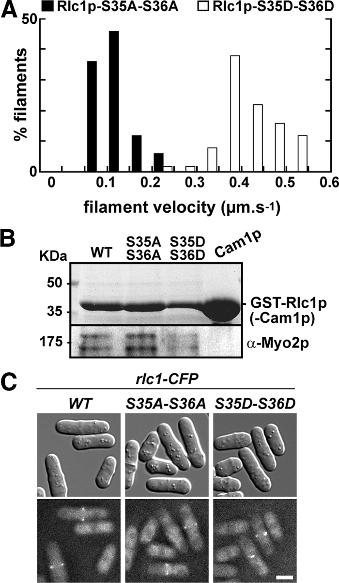
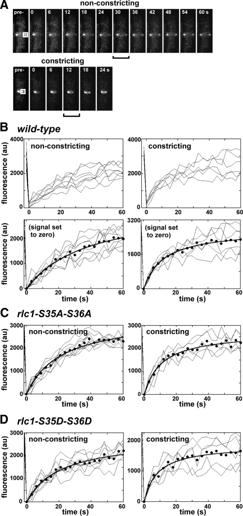
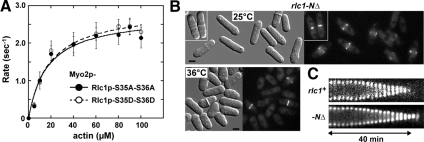
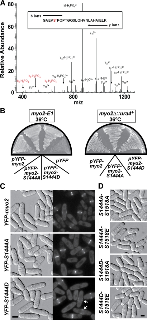
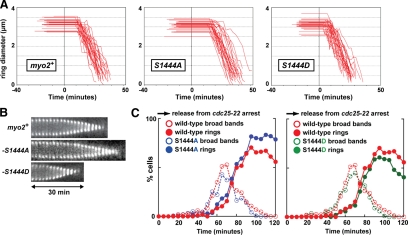
We investigated the role of regulatory light-chain (Rlc1p) and heavy-chain phosphorylation in controlling fission yeast myosin-II (Myo2p) motor activity and function during cytokinesis. Phosphorylation of Rlc1p leads to a fourfold increase in Myo2p's in vitro motility rate, which ensures effective contractile ring constriction and function. Surprisingly, unlike with smooth muscle and nonmuscle myosin-II, RLC phosphorylation does not influence the actin-activated ATPase activity of Myo2p. A truncated form of Rlc1p lacking its extended N-terminal regulatory region (including phosphorylation sites) supported maximal Myo2p in vitro motility rates and normal contractile ring function. Thus, the unphosphorylated N-terminal extension of Rlc1p can uncouple the ATPase and motility activities of Myo2p. We confirmed the identity of one out of two putative heavy-chain phosphorylation sites previously reported to control Myo2p function and cytokinesis. Although in vitro studies indicated that phosphorylation at Ser-1444 is not needed for Myo2p motor activity, phosphorylation at this site promotes the initiation of contractile ring constriction.


Figures

Figure 1.
Figure 1.
Phosphorylation of Rlc1p speeds up the in vitro motility rate of Myo2p. (A) Epifluorescence microscopy was used to generate time-lapse movies tracking movement of rhodamine-labeled actin filaments across the surface of Myo2p-coated coverslips. Myo2p samples were one-step purified. The histogram charts the distribution of Myo2p-driven actin filament gliding rates for preparations containing an unphosphorylatable form of Rlc1p (AA, ■) or a phospho-mimicking form (DD, □). Rates were derived from independent experiments using three separate preparations of Myo2p (n = 60 filaments). See Supplementary Movie 1. (B) Myo2p copurifies with both Rlc1p-AA and -DD isolated from cell extracts. Top panel, a Coomassie-stained gel indicating isolation and enrichment of wild-type GST-Rlc1p (positive control), GST-tagged mutants, and GST-Cam1p/calmodulin (negative control). Bottom panel, a Western blot indicating copurification of endogenous Myo2p with all three Rlc1p forms. (C) Wild-type, AA, and DD forms of Rlc1p-CFP localize to the contractile ring. Fusion proteins were visualized using fluorescence microscopy. Top panels, DIC images; bottom panels, the corresponding CFP images. Bar, 4 μm.
Figure 2.
Figure 2.
Phosphorylation of Rlc1p promotes contractile ring integrity and cytokinesis. (A) Cell morphology and contractile rings viewed by DIC and epifluorescence imaging of a chromosomal YFP-Myo2p fusion are shown for rlc1-AA (left panels) and rlc1-DD (right panels) strains. Cells were grown in rich YE5S media at either 25°C (top) or 36°C (bottom) before imaging. Bars, 4 μm. (B) rlc1+, rlc1-AA, rlc1-DD, and rlc1-NΔ strains grown in YE5S media at 25, 32, and 36°C were treated with Hoechst stain to mark nuclei. The plot summarizes the cytokinesis phenotypes of cells quantitated by scoring multinucleate cells (3+ nuclei/cell) using fluorescence microscopy (n = 300 cells). (C) The ability of wild-type, rlc1-AA, and rlc1-DD cells to grow at 32°C on a YE5S plate containing 1 M KCl are compared.
Figure 3.
Figure 3.
Effect of Rlc1p phosphorylation on contractile ring dynamics. (A) Plots charting the assembly, dwell, and constriction phases of individual rings from rlc1+ (n = 30), rlc1-AA (n = 25), rlc1-DD (n = 30), and rlc1-NΔ (n = 30) cells recorded using time-lapse fluorescent microscopy of YFP-Myo2p. Only rlc1-AA cells that successfully completed constriction were included in the analysis. For each plot the length of flat-line traces shown at negative time represent assembly time, whereas the length of flat line traces shown at positive time correspond to the dwell time. Diagonal lines correspond to the constriction phase, derived from measuring ring diameter over time for each cell. Cells were grown in YE5S media at 25°C and imaged by time-lapse epifluorescence microscopy (at 23°C). The schematic of a fission yeast cell (shown above the rlc1+ plot) illustrates the timing of contractile ring (red) assembly, dwell, and constriction phases as they relate to the division and position of nuclei (gray) during mitosis. (B) Kymographs comparing YFP-Myo2p contractile ring dynamics of rlc1+, rlc1-AA, and rlc1-DD cells. Each kymograph is made up of a series of thin slices centered on the contractile ring using images captured every 2 min. Slice height: 4.4 μm. Kymographs start from the point at which rings have assembled, spanning dwell and constriction phases. Kymographs are aligned based on the time point at which constriction begins (indicated by the arrow). See Supplementary Movie 2. (C) Additional kymographs comparing contractile ring dynamics from rlc1-AA and -DD cells during the constriction phase. (D) Kymograph of a contractile ring from an rlc1-AA cell that fails to complete constriction.
Figure 4.
Figure 4.
Phosphorylation of Rlc1p promotes Myo2p function in vivo. The phenotypes of rlc1+, rlc1-AA, and rlc1-DD strains lacking the nonessential myosin-II (Myp2p) were examined. (A) Representative fields of cells imaged by DIC microscopy following growth in YE5S media at 25 and 29°C. Bar, 4 μm. (B) Cells grown at 25 or 29°C were treated with Hoechst stain to mark nuclei. The plot summarizes the cytokinesis phenotypes of cells measured by scoring multinucleate cells using fluorescence microscopy (n = 300).
Figure 5.
Figure 5.
The rate of Myo2p exchange in the contractile ring increases during constriction. FRAP was used to measure YFP-Myo2p exchange rates in nonconstricting and constricting contractile rings using confocal laser scanning fluorescence microscopy. Cells were grown in YE5S media at 25°C before imaging at ambient temperature. (A) Micrographs charting recovery of YFP-Myo2p fluorescence in nonconstricting (top) and constricting (bottom) rings from representative rlc1+ cells. Panels on the far left show prebleached rings that were subsequently bleached at a region of interest (ROI, white box). Subsequent panels chart recovery of signal at the ring (0–60 s). Brackets indicate the point when recovery is half-maximal (t1/2). Panel width: 5 μm. (B–D) Plots charting fluorescence intensity versus time for ROIs on nonconstricting and constricting rings from rlc1+, rlc1-AA, and rlc1-DD cells. Fluorescence intensities measured before (−1.5 s) and after photobleaching (every 3 s, 0–60 s) are plotted. Individual ROI traces (thin lines) are shown (n = 5–10) along with an average fit (●, thick line). Datasets for each trace were corrected for additional bleaching encountered during time-lapse imaging by a control ROI (derived from an unbleached ring in the same field of cells). To facilitate curve fitting, zero signal intensity was set for each trace by subtracting residual YFP-Myo2p signal (detected at the ROI at the first time point after bleach, 0 s) from all trace values. Examples of datasets lacking this correction are provided for experiments with the rlc1+ strain (B, top panels).
Figure 6.
Figure 6.
The N-terminal extension of Rlc1p, but not Myo2p actin-activated ATPase activity, accounts for the reduced in vitro motility rate of dephosphorylated Rlc1p. (A) Actin-activated Mg2+-ATPase activity of two-step purified Myo2p as a function of actin concentration. The two curves represent Myo2p-Cdc4p associated with Rlc1p-AA (●) and Rlc1p-DD (○). Basal Myo2p ATPase activities (detected from control reactions lacking actin) and background Pi from actin (determined from controls lacking myosin) were subtracted from all measurements taken in the presence of 0–100 μM actin filaments. Each curve was generated from average values obtained from four different datasets and fit to Michaelis-Menten kinetics using KaleidaGraph software. (B) Cell morphology and contractile rings viewed by DIC and epifluorescence imaging of YFP-Myo2p are shown for the rlc1-NΔ strain in which Rlc1p lacks its regulatory N-terminus (amino acids 3–36). Cells were grown in rich media at either 25 or 36°C before imaging. Bar, 4 μm. (C) Representative kymographs comparing contractile ring dynamics of YFP-Myo2p in rlc1+ and rlc1-NΔ cells. Cells were prepared and kymographs generated from time-lapse fluorescence microscopy images as detailed in Figure 3B. Dwell and constriction phases are charted upon completion of ring assembly.
Figure 7.
Figure 7.
Cytokinesis is not dependent on the phosphorylation state of the Myo2p tail at Ser-1444 or -1518. (A) Electrospray ionization liquid chromatography ion trap mass spectrometry was performed on the Myo2p heavy chain. The Coomassie-stained heavy-chain band was cut out of an SDS-PAGE gel and analyzed. The phosphorylated peptide GAEVS*PQPTGQSLQHVNLAHAIELK produced a precursor ion at m/z 902.35 (z = 3) and was selected for MS/MS fragmentation. The largest MS/MS ion, m/z 869.93 (z = 3) corresponded to the precursor ion with the neutral loss of phosphate (−97.97 Da). The second and third largest MS/MS ions m/z 978.84 and 1091.83 (z = 2), corresponded to fragment ions y18 and y20, respectively. These ions are generated from cleavage of the precursor ion on the N-terminal side of Pro1445 or Pro1447, respectively, and their high abundance is anticipated (Breci et al., 2003). The MS/MS ions m/z 426.09, 523.22, and 651.21 (z = 1) correspond to fragment ions b5, b6, and b7 with the neutral loss of phosphate. These fragment ions indicate phosphorylation is located on Ser-1444 rather than Ser-1451. (B) pYFP-myo2-S1444A and -S1444D complement the temperature-sensitive lethality of the myo2-E1 mutant (left plate) and rescue growth of a myo2Δ strain (right plate). pYFP-myo2: positive control; pYFP: negative control lacking a myo2 insert. Cells were streaked out and grown at 36°C on EMM minimal media plates lacking leucine. (C) Gene replacement of myo2 with YFP-myo2-S1444A or -S1444D forms does not impact cytokinesis (left panels, DIC). Localization of integrated YFP-Myo2p, YFP-Myo2p-S1444A, and YFP-Myo2p-S1444D was captured by epifluorescence microscopy (right panels). Arrows in the bottom right panel highlight the presence of YFP-Myo2p-S1444D in the broad bands of assembling contractile rings. Bar, 4 μm. (D) Morphologies of YFP-myo2-S1444A-S1518A, YFP-myo2-S1444A-S1518E, YFP-myo2-S1444D-S1518A, and YFP-myo2-S1444D-S1518E double mutants. All cells were grown at 32°C in YE5S media before imaging. Bar, 4 μm.
Figure 8.
Figure 8.
Phosphorylation of the Myo2p tail at Ser-1444 favors the initiation of ring constriction. (A) Plots charting the assembly, dwell, and constriction phases of individual rings from YFP-myo2, YFP-myo2-S1444A, and YFP-myo2-S1444D cells (n = 30; prepared as described in Figure 3A). (B) Representative kymographs generated from YFP-myo2, YFP-myo2-S1444A, and YFP-myo2-S1444D cells are displayed from the point at which rings have compacted from assembling broad bands. Cells were prepared and kymographs generated as detailed in Figure 3B. Nonconstricting and constricting phases are compared. (C) Time courses of broad band (assembling rings) and contractile ring appearance in cdc25-22 cells harboring a YFP-myo2, YFP-myo2-S1444A, or YFP-myo2-S1444D fusion. Cells were grown up in YE5S media at 25°C and then arrested in the G2 phase of the cell cycle by shifting cultures to 36°C (utilizing the temperature-sensitive cdc25-22 mutation). Synchronized cells were released into mitosis by growth at 25°C (time zero) and cell samples collected every 5 min from 0 to 120 min. The percentage of cells possessing broad bands or fully formed rings of YFP-Myo2p were scored using epifluorescence microscopy analysis. Plots for S1444A (left, blue circles) and S1444D (right, green circles) cells are overlaid on the wild-type time course. (For each strain n = 120–200 cells/time point).

Similar articles

Cited by

References

    1. Andreev O. A., Saraswat L. D., Lowey S., Slaughter C., Borejdo J. Interaction of the N-terminus of chicken skeletal essential light chain 1 with F-actin. Biochemistry. 1999;38:2480–2485. - PubMed
    1. Bahler J., Wu J. Q., Longtine M. S., Shah N. G., McKenzie A., 3rd, Steever A. B., Wach A., Philippsen P., Pringle J. R. Heterologous modules for efficient and versatile PCR-based gene targeting in Schizosaccharomyces pombe. Yeast. 1998;14:943–951. - PubMed
    1. Balasubramanian M. K., McCollum D., Chang L., Wong K. C., Naqvi N. I., He X., Sazer S., Gould K. L. Isolation and characterization of new fission yeast cytokinesis mutants. Genetics. 1998;149:1265–1275. - PMC - PubMed
    1. Bezanilla M., Forsburg S. L., Pollard T. D. Identification of a second myosin-II in Schizosaccharomyces pombe: Myp2p is conditionally required for cytokinesis. Mol. Biol. Cell. 1997;8:2693–2705. - PMC - PubMed
    1. Bezanilla M., Wilson J. M., Pollard T. D. Fission yeast myosin-II isoforms assemble into contractile rings at distinct times during mitosis. Curr. Biol. 2000;10:397–400. - PubMed

Publication types

MeSH terms