. 2009 Jul 21;106(29):12189-94.
doi: 10.1073/pnas.0810332106. Epub 2009 Jul 1.

Control of heart rate by cAMP sensitivity of HCN channels

Affiliations

Control of heart rate by cAMP sensitivity of HCN channels

Jacqueline Alig et al. Proc Natl Acad Sci U S A. .

Abstract

"Pacemaker" f-channels mediating the hyperpolarization-activated nonselective cation current I(f) are directly regulated by cAMP. Accordingly, the activity of f-channels increases when cellular cAMP levels are elevated (e.g., during sympathetic stimulation) and decreases when they are reduced (e.g., during vagal stimulation). Although these biophysical properties seem to make f-channels ideal molecular targets for heart rate regulation by the autonomic nervous system, the exact contribution of the major I(f)-mediating cardiac isoforms HCN2 and HCN4 to sinoatrial node (SAN) function remains highly controversial. To directly investigate the role of cAMP-dependent regulation of hyperpolarization activated cyclic nucleotide activated (HCN) channels in SAN activity, we generated mice with heart-specific and inducible expression of a human HCN4 mutation (573X) that abolishes the cAMP-dependent regulation of HCN channels. We found that hHCN4-573X expression causes elimination of the cAMP sensitivity of I(f) and decreases the maximum firing rates of SAN pacemaker cells. In conscious mice, hHCN4-573X expression leads to a marked reduction in heart rate at rest and during exercise. Despite the complete loss of cAMP sensitivity of I(f), the relative extent of SAN cell frequency and heart rate regulation are preserved. Our data demonstrate that cAMP-mediated regulation of I(f) determines basal and maximal heart rates but does not play an indispensable role in heart rate adaptation during physical activity. Our data also reveal the pathophysiologic mechanism of hHCN4-573X-linked SAN dysfunction in humans.


Conflict of interest statement

The authors declare no conflict of interest.

Figures

Fig. 1.
Fig. 1.
Generation and characterization of transgenic mice. (A) Schematic illustration of the Tet-Off system. (B) Fluorescence microscopic images (EGFP autofluorescence) of the SAN region of an αMHC-tTA–driven EGFP indicator line (20). (C) Northern blot detection of hHCN4–573X–specific RNA in total cardiac RNA samples from offspring of 2 independent founder lines (A and F) revealing 2 different expression levels. Total RNA isolated from a wild-type mouse heart was used as a negative control (“−”). In vitro–synthesized hHCN4 cRNA (100 pg) was added to the control RNA as a positive control (“+”). (D) Western blot detection of transgenic hHCN4–573X proteins in total heart tissue lysates from double-transgenic (mutant) offspring of both founder lines and from a nontransgenic control mouse (“−”). Total lysates of human embryonic kidney (HEK-293) cells transiently transfected with hHCN4–573X were used as a positive control (12). (E) Region-specific Western blot analysis of proteins isolated from the right atrium (RA), left atrium (LA), right ventricle (RV), and left ventricle (LV). hHCN4–573X protein was detectable in atrial tissue lysates from both founder lines, whereas ventricular transgene expression was detected only in line F. (F) DOX-dependent regulation of hHCN4–573X expression and comparison with endogenous mHCN4 protein levels analyzed by Western blot. Total heart lysates were obtained from mutants on DOX (“D”) and from DOX-water (“DW”) animals. Abbreviations: tTA, Tet-Off system transactivator; Tre, tetracycline-responsive element; CT, crista terminalis, SVC, superior vena cava.
Fig. 2.
Fig. 2.
β-adrenergic regulation of If in control and mutant SAN cells. Black indicates recording under control conditions with normal Tyrode's solution (Tyr); red indicates data obtained after ISO stimulation. (A and B) Examples of representative voltage-clamp recordings of hyperpolarization-induced currents from acutely isolated control (A) or mutant (B; founder line A) SAN pacemaker cells (protocol shown below traces). (C and D) Sample traces of control (C) and mutant If (D) in the absence or presence of 100 nM ISO. (E and F) Current density–voltage relationships at baseline revealing comparable If densities in control (black squares) and mutant cells (black circles). Stimulation with 100 nM ISO increased If density in control cells (E, red squares), but not in mutant cells (F, red circles). (G) If activation curves in control (black squares) and mutant cells (black circles) (26). If in mutant cells activated at more negative voltages than in control cells. In control cells, application of ISO caused a significant shift in the If activation curve to more positive voltages (red squares). In contrast, no significant ISO-dependent shift was observed in mutant cells (red circles). (H) Analysis of β-adrenergic regulation of ICa,L in control and mutant cells. The bar graph shows current–time integrals of ICa,L. ICa,L obtained from the same set of experiments shown in (C–F). ICa,L was activated by depolarizing voltage steps from a holding potential of −60 mV to −35 mV. At this test voltage, ICa,L is generated predominantly by Cav1.3 channels (29). ICa,L was recorded in normal Tyrode's solution (Tyr) at 35 °C with and without 100 nM ISO. ICa,L in control (n = 5; filled bars) and mutant SAN cells (n = 5; open bars) was similarly stimulated by application of ISO. ICa,L waveform was integrated over a time interval of 60 ms starting from the current onset (30). *P < .05.
Fig. 3.
Fig. 3.
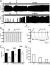
Pacemaker activity in SAN cells of control and mutant mice. (A) Fast and regular firing was observed in SAN cells of control mice (Top), whereas most mutant (founder line A) SAN cells showed only intermittent action potential firing, alternating with oscillations in membrane voltage (Bottom). Activation of the β-adrenergic receptor by a submaximal dose of ISO (2 nM) restored regular firing in mutant cells, although at a slower rate than in control cells. (B) Fast-time scale recording of pacemaker activity of a control cell (Left) and a mutant cell (Right). Control cells showed a regular firing rate in Tyrode's solution (Tyr; black line) and accelerated firing in ISO (gray line). In most mutant cells, ISO application restored regular firing activity, but the maximum rate was still slower than that in control cells. (C) Pacemaker activity in mutant cells (n = 10; open bars) was increased by stimulation with 2 nM or 100 nM ISO, but the cell firing rate at each dose tested did not reach that of control cells (n = 12; filled bars). Note that the firing rate of mutant cells was reduced at baseline (Tyr) and at each ISO concentration tested (Table S1). (D) The percentage of quiescent pacemaker cells decreased in a dose-dependent manner on superfusion with 2 nM or 100 nM ISO. ***P < .001.)
Fig. 4.
Fig. 4.
The selective If inhibitor IVA reduced the heart rate in control mice, but not in mutant mice. (A) Application of 3 or 10 μM IVA onto acutely isolated control (filled bars) or mutant (open bars) SAN cells revealed similar If inhibition at a membrane potential of −105 mV. (B and C) Mean heart rate in control mice (B; n = 3) and mutant mice (C; n = 4). Each animal received an i.p. injection of physiological saline (0.9% NaCl), followed by IVA at a dose of 3 mg/kg (3 IVA) or 6 mg/kg (6 IVA) at 72-h intervals, to allow washout of the drug. The heart rate was measured for 3 h before injection of either NaCl or IVA and then for 3 h after injection. Note the reduced basal heart rate in the mutant mice. The aforementioned periods for heart rate averaging were selected because IVA reached its maximum effect 2 h after injection of 3 mg/kg and 1 h after injection of 6 mg/kg (data not shown). Data were compared using ANOVA and the Newman–Keuls posthoc test. *P < .05.
Fig. 5.
Fig. 5.
Activity-dependent regulation of heart rate in control and mutant mice. (A) Experimental protocol. Mice were raised on DOX and implanted with telemetric devices. ECG recordings were obtained from the same animals raised on DOX (B), after withdrawal of DOX (water) (C), and during atropine and propranolol (A/P) administration (D). (B) Heart rate increased depending on the level of spontaneous home cage activity of control animals (filled bars; n = 4) and mutant animals on DOX (open bars; n = 6). (C) When hHCN4–573X expression was induced by DOX withdrawal (“on water”), the control mice exhibited the same activity-dependent heart rate as those raised on DOX. The mutant mice on water had a significantly lower heart rate at all activity levels. (D) Heart rates of controls and mutants during autonomic nervous system blockade with A/P. (E) Range of heart rate regulation calculated from the differences between maximum and minimum heart rates in (B–D). (F and G) Histogram of normalized average heart rates recorded during a 24-h period from control (F) and mutant (G; line A) mice on DOX (black), on water (red), and during autonomic nervous system blockade with A/P (blue). (H) Sample ECG raw traces (epochs of 2 s) from control and mutant animals on DOX or water at low and high physical activity levels showing reduced heart rate in mutants, but no signs of SAN dysfunction, conductance abnormalities, or ventricular arrhythmias. For quantification of PQ and QTc interval durations, see Fig. S3. (Scale bars: 0.2 mV.) ***P < .001.

References

    1. Lamas GA, et al. The mode selection trial (MOST) in sinus node dysfunction: Design, rationale, and baseline characteristics of the first 1000 patients. Am Heart J. 2000;140:541–551. - PubMed
    1. Gillman MW, Kannel WB, Belanger A, D'Agostino RB. Influence of heart rate on mortality among persons with hypertension: The Framingham Study. Am Heart J. 1993;125:1148–1154. - PubMed
    1. Fox K, et al. Resting heart rate in cardiovascular disease. J Am Coll Cardiol. 2007;50:823–830. - PubMed
    1. Maltsev VA, Lakatta EG. Dynamic interactions of an intracellular Ca2+ clock and membrane ion channel clock underlie robust initiation and regulation of cardiac pacemaker function. Cardiovasc Res. 2008;77:274–284. - PubMed
    1. Mangoni M, Nargeot J. Genesis and regulation of the heart automaticity. Physiol Rev. 2008;88:919–982. - PubMed

Publication types

MeSH terms