. 2009 Jul 1;29(26):8474-82.
doi: 10.1523/JNEUROSCI.0378-09.2009.

Sustained conditioned responses in prelimbic prefrontal neurons are correlated with fear expression and extinction failure

Affiliations

Sustained conditioned responses in prelimbic prefrontal neurons are correlated with fear expression and extinction failure

Anthony Burgos-Robles et al. J Neurosci. .

Abstract

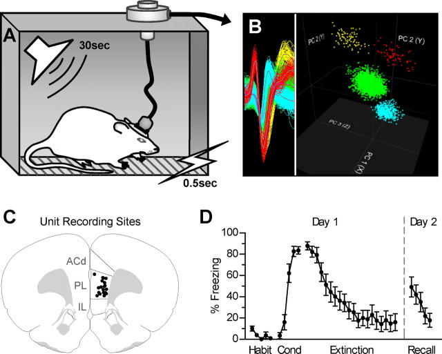
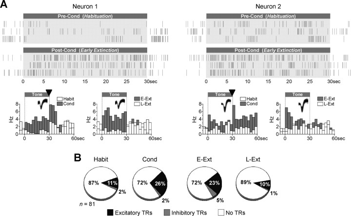
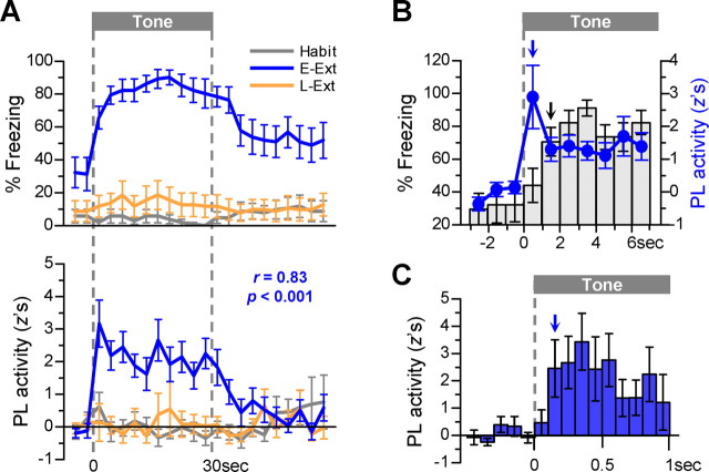
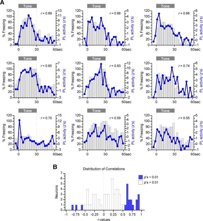
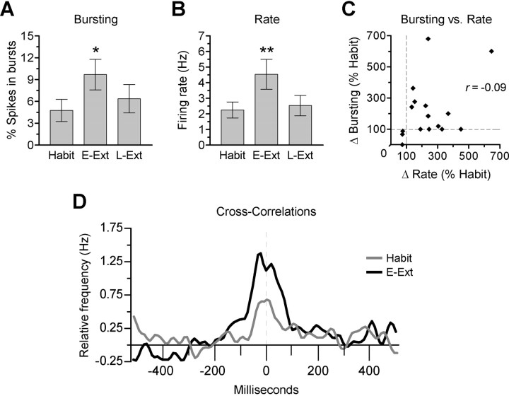
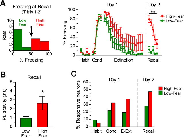
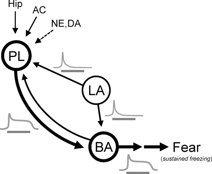
During auditory fear conditioning, it is well established that lateral amygdala (LA) neurons potentiate their response to the tone conditioned stimulus, and that this potentiation is required for conditioned fear behavior. Conditioned tone responses in LA, however, last only a few hundred milliseconds and cannot be responsible for sustained fear responses to a tone lasting tens of seconds. Recent evidence from inactivation and stimulation studies suggests that the prelimbic (PL) prefrontal cortex is necessary for expression of learned fears, but the timing of PL tone responses and correlations with fear behavior have not been studied. Using multichannel unit recording techniques in behaving rats, we observed sustained conditioned tone responses in PL that were correlated with freezing behavior on a second-to-second basis during the presentation of a 30 s tone. PL tone responses were also correlated with conditioned freezing across different experimental phases (habituation, conditioning, extinction). Moreover, the persistence of PL responses after extinction training was associated with failure to express extinction memory. Together with previous inactivation findings, the present results suggest that PL transforms transient amygdala inputs to a sustained output that drives conditioned fear responses and gates the expression of extinction. Given the relatively long latency of conditioned responses we observed in PL (approximately 100 ms after tone onset), we propose that PL integrates inputs from the amygdala, hippocampus, and other cortical sources to regulate the expression of fear memories.


Figures

Figure 1.
Figure 1.
Multichannel unit recordings were performed in the prelimbic subregion of the medial prefrontal cortex of rats undergoing auditory fear conditioning and extinction. A, Rats were fear conditioned with tones of 30 s in length that coterminated with brief footshocks. Extinction training was conducted 2 h after conditioning, and the recall test occurred 24 h after extinction. B, Representative extracellular waveform sorting. The image shows superimposed waveforms of four simultaneously recorded neurons and the clusters they formed in three-dimensional space after applying principal component analysis. C, Coronal drawing shows location of recording electrodes [bregma +2.80 mm; adapted from Swanson (1992); ACd, dorsal anterior cingulate]. D, Percentage freezing to the tone (n = 17). Significant acquisition and extinction were observed on day 1, and substantial recall of extinction was observed on day 2. In this and subsequent figures, error bars illustrate SEM; Habit indicates habituation, and Cond indicates conditioning.
Figure 2.
Figure 2.
Prelimbic neurons showed sustained excitatory responses to conditioned tones. A, Spike trains of two representative neurons before conditioning and after fear conditioning. Perievent time histograms show the response of these two neurons to tones across different phases of the experiment (bin width, 3 s). These neurons responded to tones in a sustained manner during high-fear states (conditioning and early extinction) and stopped responding to tones by the end of the extinction session (late extinction). These neurons also responded to footshocks (▾) during the conditioning phase. B, Pie charts illustrate the percentage of PL neurons showing excitatory or inhibitory tone responses (TRs; see Materials and Methods for criteria used to detect tone responses). The proportion of neurons showing excitatory tone responses significantly increased from habituation to conditioning and decreased from early extinction to late extinction (χ2 test, p's < 0.05). A small proportion of neurons showed inhibitory tone responses, which did not change significantly with conditioning or extinction training (p's > 0.27). In this and subsequent figures, E-Ext indicates early extinction, and L-Ext indicates late extinction.
Figure 3.
Figure 3.
Conditioned responses in prelimbic neurons were highly correlated with the time course of conditioned freezing. A, Top, Freezing was assessed on a second-to-second time scale and plotted in 3 s bins. A, Bottom, Average tone response of neurons classified as tone responsive in early extinction (n = 19 of 81, 23%). Neuronal activity resembled freezing in all phases, especially during early extinction in which there was a significant correlation between the two measures across bins (r = 0.83). B, Response latency of PL activity (blue line plot) and freezing (bars) during early extinction (1 s bins are plotted). Significant tone responses in PL were evident from the very first second after tone onset, whereas significant freezing was not observed until after two seconds (↓ p < 0.001, compared with pretone). C, Earliest tone response latency in PL. Short-latency responses were examined in the 15 neurons showing significant z-scores in the first 3 s bin in A. These neurons did not start signaling the tone until 100 ms after tone onset (↓ p < 0.05, compared with pretone).
Figure 4.
Figure 4.
Single-neuron examples of prelimbic activity versus freezing. A, The activity of single neurons was averaged over five trials during early extinction, and the normalized tone response (line plots) was graphed on top of freezing values for a given rat (bars). Nine examples of activity–behavior correlations are shown in descending order. B, Distribution of 67 correlations obtained (14 of 81 neurons were excluded from this analysis because of low firing rates, <0.05 Hz). There was a greater number of positive correlations than negative correlations (66 vs 34%; binomial proportions test, p = 0.007). Furthermore, although 24% of the correlations were significantly positive (16 of 67, blue bars on the right), only 4% were significantly negative (3 of 67, blue bars on the left).
Figure 5.
Figure 5.
Conditioning increased bursting and synchrony in prelimbic neurons. A, B, Average bursting and rate during tone periods of neurons that were tone-responsive during early extinction (n = 19). Bursting was significantly higher during early extinction than during habituation (*p = 0.03) and tended to reduce with extinction training (p = 0.19, early ext vs late ext). B, These training-induced changes in bursting are similar to those observed in firing rate (**p's < 0.01, early ext vs habit and late ext). C, Relationship between rate and bursting during early extinction. Each dot represents a single neuron, and values are expressed as changes from habituation levels. The lack of correlation (r = −0.09) suggests that changes in bursting occurred independently of changes in firing rate. D, Cross-correlations between simultaneously recorded cell pairs during tone periods. Histograms show the average of 36 of 147 (24%) correlograms showing significant peaks during early extinction (see Materials and Methods for statistical analysis). The peak of the group correlogram was significantly higher during early extinction than during habituation (paired t test, p = 0.011). (Correlograms were smoothed using Gaussian filter, with filter width of 3 10 ms bins; y-axis represents relative firing frequency after subtracting shift-predictor values.)
Figure 6.
Figure 6.
Failure to recall extinction was associated with increased prelimbic tone responses. A, Rats were divided into two subgroups based on the degree of extinction recall: rats showing good extinction recall (low-fear, <50% freezing in the first two trials; n = 8) and rats showing poor extinction recall (high-fear, >50% freezing; n = 7; **p's <0.01). No significant group differences in freezing were detected in conditioning and extinction training. B, Magnitude of tone responses at the recall phase in the two subgroups (9 tone-responsive neurons in each subgroup). High-fear rats showed larger PL tone responses at recall than low-fear rats, suggesting that PL activity contributed to impaired extinction recall (*p = 0.046). C, Percentage of tone-responsive neurons at different phases of the experiment. Only neurons maintained throughout all phases were considered for this analysis (low-fear, n = 32; high-fear, n = 19). Compared with low-fear rats, high-fear rats showed a higher percentage of tone-responsive neurons at recall, as well as during conditioning and early extinction.
Figure 7.
Figure 7.
Suggested model by which the prelimbic cortex maintains the expression of conditioned freezing. PL receives convergent inputs from several sources: the LA and BA (providing information regarding the tone–shock association), the hippocampus (Hip) (providing contextual information), and the auditory cortex (AC) (providing auditory information). By integrating these inputs, together with fear-induced release of neuromodulators such as norepinephrine (NE) and dopamine (DA), PL could convert phasic inputs from the amygdala into sustained outputs (see traces in gray). In this scenario, thus, PL integrates auditory, contextual, and stress-related signals to calculate an appropriate behavioral response to a conditioned stimulus.

Comment in

References

    1. Amorapanth P, Nader K, LeDoux JE. Lesions of periaqueductal gray dissociate-conditioned freezing from conditioned suppression behavior in rats. Learn Mem. 1999;6:491–499. - PMC - PubMed
    1. Anglada-Figueroa D, Quirk GJ. Lesions of the basal amygdala block expression of conditioned fear but not extinction. J Neurosci. 2005;25:9680–9685. - PMC - PubMed
    1. Baeg EH, Kim YB, Jang J, Kim HT, Mook-Jung I, Jung MW. Fast spiking and regular spiking neural correlates of fear conditioning in the medial prefrontal cortex of the rat. Cereb Cortex. 2001;11:441–451. - PubMed
    1. Barrett D, Shumake J, Jones D, Gonzalez-Lima F. Metabolic mapping of mouse brain activity after extinction of a conditioned emotional response. J Neurosci. 2003;23:5740–5749. - PMC - PubMed
    1. Blanchard DC, Blanchard RJ. Innate and conditioned reactions to threat in rats with amygdaloid lesions. J Comp Physiol Psychol. 1972;81:281–290. - PubMed

Publication types

LinkOut - more resources