. 2009 Sep;297(3):G471-9.
doi: 10.1152/ajpgi.00012.2009. Epub 2009 Jul 1.

Enterocyte-specific epidermal growth factor prevents barrier dysfunction and improves mortality in murine peritonitis

Affiliations

Enterocyte-specific epidermal growth factor prevents barrier dysfunction and improves mortality in murine peritonitis

Jessica A Clark et al. Am J Physiol Gastrointest Liver Physiol. 2009 Sep.

Abstract

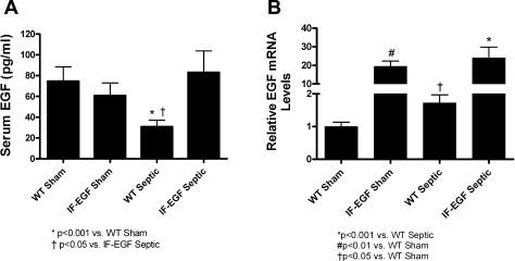
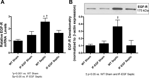
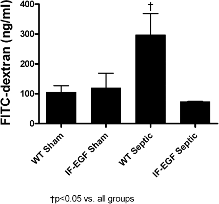
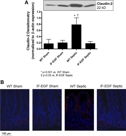
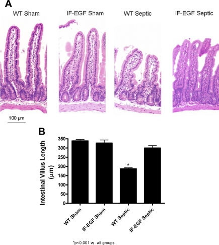
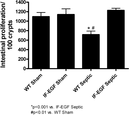
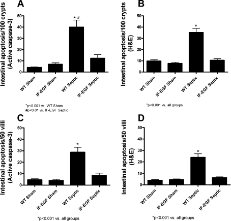
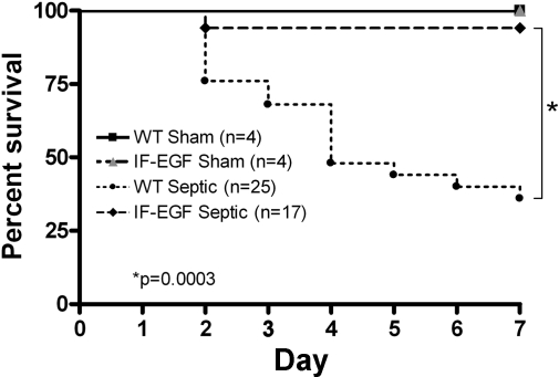
Systemic administration of epidermal growth factor (EGF) decreases mortality in a murine model of septic peritonitis. Although EGF can have direct healing effects on the intestinal mucosa, it is unknown whether the benefits of systemic EGF in peritonitis are mediated through the intestine. Here, we demonstrate that enterocyte-specific overexpression of EGF is sufficient to prevent intestinal barrier dysfunction and improve survival in peritonitis. Transgenic FVB/N mice that overexpress EGF exclusively in enterocytes (IFABP-EGF) and wild-type (WT) mice were subjected to either sham laparotomy or cecal ligation and puncture (CLP). Intestinal permeability, expression of the tight junction proteins claudins-1, -2, -3, -4, -5, -7, and -8, occludin, and zonula occludens-1; villus length; intestinal epithelial proliferation; and epithelial apoptosis were evaluated. A separate cohort of mice was followed for survival. Peritonitis induced a threefold increase in intestinal permeability in WT mice. This was associated with increased claudin-2 expression and a change in subcellular localization. Permeability decreased to basal levels in IFABP-EGF septic mice, and claudin-2 expression and localization were similar to those of sham animals. Claudin-4 expression was decreased following CLP but was not different between WT septic mice and IFABP-EGF septic mice. Peritonitis-induced decreases in villus length and proliferation and increases in apoptosis seen in WT septic mice did not occur in IFABP-EGF septic mice. IFABP-EGF mice had improved 7-day mortality compared with WT septic mice (6% vs. 64%). Since enterocyte-specific overexpression of EGF is sufficient to prevent peritonitis-induced intestinal barrier dysfunction and confers a survival advantage, the protective effects of systemic EGF in septic peritonitis appear to be mediated in an intestine-specific fashion.


Figures

Fig. 1.
Fig. 1.
Systemic and intestinal EGF levels. Circulating EGF levels (A) were significantly lower in wild-type (WT) septic than WT sham animals but were not statistically different between intestinal fatty acid binding protein (IFABP)-EGF (IF-EGF) sham and IFABP-EGF septic mice. In contrast, intestinal EGF levels (B) were higher in WT septic than WT sham animals. Whereas intestinal EGF levels were higher in IFABP-EGF mice than WT mice, levels of intestinal EGF were not statistically different between IFABP-EGF sham and IFABP-EGF septic mice.
Fig. 2.
Fig. 2.
Intestinal EGF receptor (EGF-R) expression. Relative mRNA levels (A) and protein expression (B) of EGF-R were quantified in jejunal tissue via quantitative real-time PCR and Western blot, respectively. Intestinal EGF-R mRNA and protein levels were increased in WT septic mice compared with shams, but enterocyte-specific overexpression of EGF resulted in normalization to sham levels. A representative blot for EGF-R is depicted; densitometry was determined by normalizing expression to β-actin.
Fig. 3.
Fig. 3.
Intestinal permeability. Intestinal permeability was assayed in vivo by measuring the amount of fluorescein isothiocyanate-conjugated dextran (FD-4) in plasma. WT septic mice had increased intestinal permeability compared with sham animals, whereas sepsis-induced increases in intestinal permeability were prevented in IFABP-EGF septic mice.
Fig. 4.
Fig. 4.
Expression and localization of claudin-2. Protein expression and localization of claudin-2 were evaluated by Western blot (A) and fluorescent immunohistochemistry (B). Claudin-2 protein levels were increased in WT septic mice compared with shams. In contrast, claudin-2 expression was normalized to sham levels in IFABP-EGF septic mice. A representative blot for claudin-2 is depicted; densitometry was determined by normalizing expression to β-actin. Fluorescent immunohistochemistry revealed that claudin-2 expression was increased and localized along the apical membrane of the villi in WT septic mice. Minimal staining for claudin-2 was observed in either the sham mice or IFABP-EGF septic mice. Representative images for each group are shown. Magnification ×20.
Fig. 5.
Fig. 5.
Intestinal villus length. Intestinal morphology (A) was evaluated in hematoxylin and eosin (H&E)-stained intestinal sections. WT septic animals appeared to have markedly shorter villi than sham animals. Villi appeared to be longer in IFABP-EGF septic mice compared with WT septic mice. Magnification ×20. Villus length was quantified in H&E-stained sections of jejunum (B). WT septic mice had significantly shorter villi compared with sham mice. Villus length was preserved in IFABP-EGF septic mice.
Fig. 6.
Fig. 6.
Intestinal proliferation. S-phase cells were quantified in 100 contiguous crypts by 5-bromo-2′-deoxyuridine staining. WT septic mice had significantly decreased intestinal proliferation, whereas IFABP-EGF septic mice exhibited normalization to sham levels.
Fig. 7.
Fig. 7.
Intestinal epithelial apoptosis. Intestinal apoptosis was quantified in crypt (A and B) and villus (C and D) epithelium by active caspase-3 staining and H&E staining. WT septic mice exhibited increased apoptosis compared with shams by both methods. In contrast, IFABP-EGF septic mice had similar levels of apoptosis to sham animals by both methods.
Fig. 8.
Fig. 8.
Effect of enterocyte-specific overexpression of EGF on mortality in sepsis. WT and IFABP-EGF mice were subjected to 2 × 23-gauge cecal ligation and puncture. Control animals underwent sham laparotomy. All mice were given antibiotics and followed for survival for 7 days. IFABP-EGF septic mice exhibited significantly improved survival compared with WT septic mice. All sham mice survived.

References

    1. Angus DC, Linde-Zwirble WT, Lidicker J, Clermont G, Carcillo J, Pinsky MR. Epidemiology of severe sepsis in the United States: analysis of incidence, outcome, and associated costs of care. Crit Care Med 29: 1303–1310, 2001. - PubMed
    1. Baker CC, Chaudry IH, Gaines HO, Baue AE. Evaluation of factors affecting mortality rate after sepsis in a murine cecal ligation and puncture model. Surgery 94: 331–335, 1983. - PubMed
    1. Basuroy S, Seth A, Elias B, Naren AP, Rao R. MAPK interacts with occludin and mediates EGF-induced prevention of tight junction disruption by hydrogen peroxide. Biochem J 393: 69–77, 2006. - PMC - PubMed
    1. Basuroy S, Sheth P, Mansbach CM, Rao RK. Acetaldehyde disrupts tight junctions and adherens junctions in human colonic mucosa: protection by EGF and l-glutamine. Am J Physiol Gastrointest Liver Physiol 289: G367–G375, 2005. - PubMed
    1. Berlanga J, Lodos J, Lopez-Saura P. Attenuation of internal organ damages by exogenously administered epidermal growth factor (EGF) in burned rodents. Burns 28: 435–442, 2002. - PubMed

Publication types

MeSH terms

LinkOut - more resources