. 2009 Jul 30;460(7255):647-51.
doi: 10.1038/nature08161. Epub 2009 Jul 1.

Evidence of Xist RNA-independent initiation of mouse imprinted X-chromosome inactivation

Affiliations

Evidence of Xist RNA-independent initiation of mouse imprinted X-chromosome inactivation

Sundeep Kalantry et al. Nature. .

Abstract

XX female mammals undergo transcriptional silencing of most genes on one of their two X chromosomes to equalize X-linked gene dosage with XY males in a process referred to as X-chromosome inactivation (XCI). XCI is an example of epigenetic regulation. Once enacted in individual cells of the early female embryo, XCI is stably transmitted such that most descendant cells maintain silencing of that X chromosome. In eutherian mammals, XCI is thought to be triggered by the expression of the non-coding Xist RNA from the future inactive X chromosome (Xi); Xist RNA in turn is proposed to recruit protein complexes that bring about heterochromatinization of the Xi. Here we test whether imprinted XCI, which results in preferential inactivation of the paternal X chromosome (Xp), occurs in mouse embryos inheriting an Xp lacking Xist. We find that silencing of Xp-linked genes can initiate in the absence of paternal Xist; Xist is, however, required to stabilize silencing along the Xp. Xp-linked gene silencing associated with mouse imprinted XCI, therefore, can initiate in the embryo independently of Xist RNA.


Figures
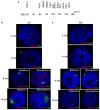
Figure 1
Figure 1. Dynamics of X-linked gene expression assayed by RNA FISH in 2-, 4-, 8-, and 16-cell wild-type (WT) and Xp-Xist1lox (Mut) female mouse embryos
a, Physical map of the eleven X-linked genes assayed. b, c, Single representative nuclei from WT and Xp-Xist1lox embryos probed for expression of G6pdx (red punctate signal) and Xist (green; in WT embryos) or Atrx (green; in Xp-Xist1lox embryos). In WT embryos, Xist is expressed exclusively from and marks the paternal X-chromosome (Xp),. In Xp-Xist1lox embryos, Atrx expression is used to mark the two Xs. In nuclei from 2- and 4-cell embryos, most genes are expressed predominantly biallelically. DNA is stained blue with 4′,6-diamidino-2-phenylindole (DAPI). d, Comparison of the distribution of nuclei displaying biallelism, monoallelism, and no signal in WT and Xp-Xist1lox embryos. n, numbers of nuclei. Only those genes/stages denoted by a ‘*’ show significant differences in the distribution of the three classes of nuclei (see Supplementary Figures 1–4).
Figure 2
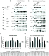
Figure 2. Dynamics of X-linked gene expression assayed by allele-specific RT-PCR in wild-type (WT) and Xp-Xist1lox (Mut) 8-16-cell mouse embryos and embryonic day 6.5 (E6.5) extra-embryonic tissues
Allele-specific RT-PCR expression analysis of the X-linked genes Xist, Ddx3x, Ube1x, Zfx, Rnf12, Atrx, Pdha1, and Utx in individual F1 hybrid WT & Xp-Xist1lox (a,c) 8-16-cell embryos (morulas) and (b,d) embryonic day 6.5 (E6.5) extra-embryonic tissues. F1 hybrid embryos were generated by a cross of the Mus molossinus (M. mol.) strain JF1 females to largely Mus domesticus (M. dom.)-derived males from wild-type and Xp-Xist1lox laboratory mice strains. Expression of the two alleles (maternal, Xm; paternal, Xp) was distinguished by single nucleotide polymorphisms between the strains resulting in a strain-specific pattern of fragments after restriction enzyme digestion (see Supplementary Methods and Supplementary Table VI). Lane 1, M. mol. allele (Xm); lane 2, M. dom. allele (Xp); lane 3, equal amounts of M. mol. and M. dom; lanes 4–6, representative F1 hybrid WT samples; lanes 7–9 representative F1 hybrid Xp-Xist1lox samples. Xist is expressed exclusively from the Xp in WT morulas and in WT E6.5 extra-embryonic tissues and is absent in Xp-Xist1lox morulas and E6.5 extra-embryonic tissues. Only those genes denoted by a ‘*’ show differences in the degree of silencing of the paternal alleles in Xp-Xist1lox morulas compared to WT embryos (see Supplementary Table I); all genes show differences in E6.5 extra-embryonic tissues (see Supplementary Table II).
Figure 3
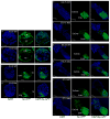
Figure 3. Expression of paternal X-linked green fluorescent protein (Xp-GFP) transgene in WT and Xp-Xist1lox (Mut) pre- and post-implantation stage female mouse embryos
a–b, Wild-type (WT) and Xp-Xist1lox blastocyst (embryonic day 3.5 [E3.5]) female embryos expressing Xp-GFP in both the trophectoderm (TE) and inner cell mass (ICM) lineages. c–d, WT and Xp-Xist1lox E4.5 female embryos silencing Xp-GFP in the TE but showing continued expression of the Xp-GFP in the ICM-derived cells. The hatched line demarcates the boundary between the TE and the ICM-derivatives. e,g,i, WT E5.75, E6.25, and E7.5 female embryos devoid of Xp-GFP expression in the extra-embryonic ectoderm and its derivatives (ExEmb; derived from the TE) due to imprinted XCI. The embryonic ectoderm (Emb), descended from the ICM, fluoresces green because of Xp-GFP expression due to random XCI that results in Xp activity in approximately half of the cells. The hatched line demarcates the boundary between the ExEmb and the Emb. f,h,j, Xp-Xist1lox E5.75, E6.25, and E7.5 female embryos with a mosaic extra-embryonic compartment that is comprised of both cells that silence and express the Xp-GFP. DNA is stained blue with DAPI.

Similar articles

Cited by

References

    1. Heard E, Chaumeil J, Masui O, Okamoto I. Mammalian X-chromosome inactivation: an epigenetics paradigm. Cold Spring Harb Symp Quant Biol. 2004;69:89–102. doi: 10.1101/sqb.2004.69.89. - DOI - PubMed
    1. Payer B, Lee JT. X chromosome dosage compensation: how mammals keep the balance. Annu Rev Genet. 2008;42:733–772. doi: 10.1146/annurev.genet.42.110807.091711. - DOI - PubMed
    1. Brown CJ, et al. A gene from the region of the human X inactivation centre is expressed exclusively from the inactive X chromosome. Nature. 1991;349:38–44. - PubMed
    1. Huynh KD, Lee JT. Inheritance of a pre-inactivated paternal X chromosome in early mouse embryos. Nature. 2003;426:857–862. - PubMed
    1. Okamoto I, Otte AP, Allis CD, Reinberg D, Heard E. Epigenetic dynamics of imprinted X inactivation during early mouse development. Science. 2004;303:644–649. - PubMed

Publication types