. 2009 Jul 23;460(7254):525-8.
doi: 10.1038/nature08156. Epub 2009 Jul 1.

Helical extension of the neuronal SNARE complex into the membrane

Affiliations

Helical extension of the neuronal SNARE complex into the membrane

Alexander Stein et al. Nature. .

Abstract

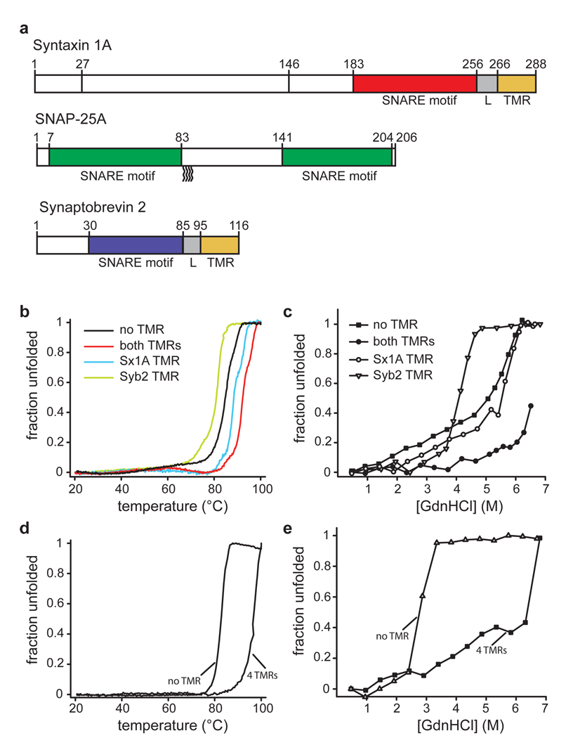
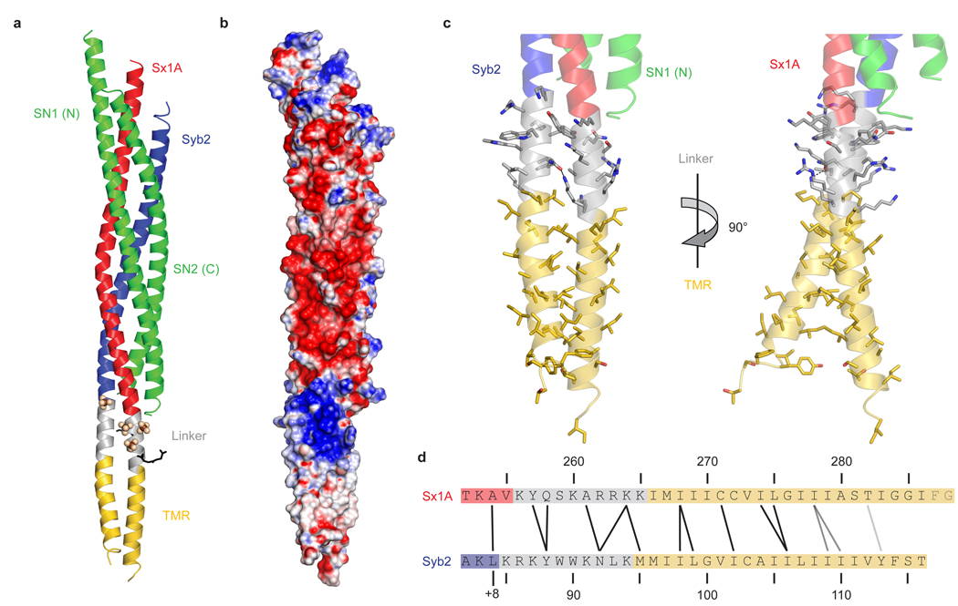
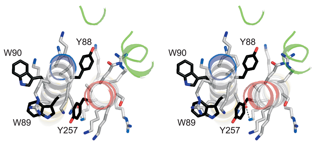
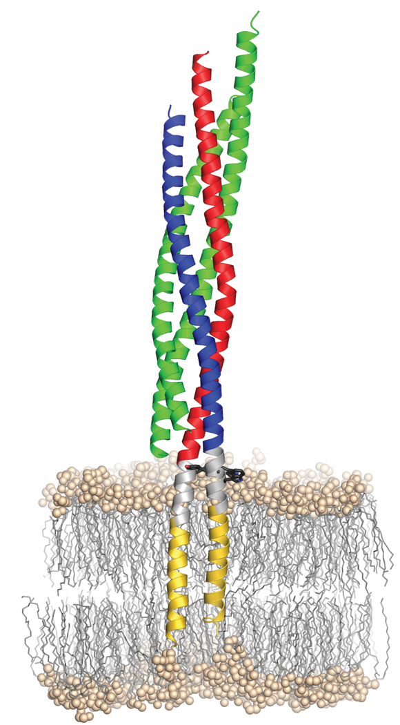
Neurotransmission relies on synaptic vesicles fusing with the membrane of nerve cells to release their neurotransmitter content into the synaptic cleft, a process requiring the assembly of several members of the SNARE (soluble N-ethylmaleimide-sensitive factor attachment protein receptor) family. SNAREs represent an evolutionarily conserved protein family that mediates membrane fusion in the secretory and endocytic pathways of eukaryotic cells. On membrane contact, these proteins assemble in trans between the membranes as a bundle of four alpha-helices, with the energy released during assembly being thought to drive fusion. However, it is unclear how the energy is transferred to the membranes and whether assembly is conformationally linked to fusion. Here, we report the X-ray structure of the neuronal SNARE complex, consisting of rat syntaxin 1A, SNAP-25 and synaptobrevin 2, with the carboxy-terminal linkers and transmembrane regions at 3.4 A resolution. The structure shows that assembly proceeds beyond the already known core SNARE complex, resulting in a continuous helical bundle that is further stabilized by side-chain interactions in the linker region. Our results suggest that the final phase of SNARE assembly is directly coupled to membrane merger.


Figures

Figure 1
Figure 1
Linkers and transmembrane regions add stability to SNARE complexes. a, Protein fragments used in this study. The complex used for crystallization contained all colored segments. For CD measurements, complexes were formed with the same syntaxin 1A fragment but with full-length synaptobrevin 2 and SNAP-25a (all cysteines replaced by serines). b–e, Unfolding of SNARE complexes, monitored by CD spectroscopy at 222nm. b, Thermal unfolding of synaptic SNARE complexes in which the TMRs were either present or in which one or both of the TMRs were lacking. Note that the complex used for crystallization unfolded at approximately 97 °C (see table 1). Sx1a, syntaxin 1A; Syb2, synaptobrevin 2. c, Unfolding of the same complexes at increasing concentrations of guanidine hydrochloride (GdnHCl). d, Thermal unfolding of endosomal SNARE complexes consisting of syntaxin 7, vti1b, syntaxin 8, and endobrevin, either containing or lacking its four TMRs. e, Unfolding of the endosomal complexes at increasing concentrations of GdnHCl.
Figure 2
Figure 2
Synaptobrevin 2 and syntaxin 1A form continuous helices. a, Ribbon plot of the synaptic SNARE complex including linkers and TMRs (colour coding as in Fig. 1a). Sulfate ions and two glycylglycylglycine molecules are depicted as spheres and black sticks, respectively. b, Surface plot showing the electrostatic potential of the synaptic SNARE complex (blue, positive charge; red, negative charge). The electrostatic surface was contoured between – 16 kT/e and + 16 kT/e. c, Ribbon plot showing linkers and TMRs of synaptobrevin 2 and syntaxin 1A. Side chains are shown as sticks, with carbons coloured as the corresponding backbone; oxygen, red; nitrogen, blue. The hydrogen-bond between Asn92 in synaptobrevin 2 and Lys264 in syntaxin 1A is depicted as a black dashed line. The right panel is rotated by 90° about the vertical axis as indicated. d, Amino acids forming interactions between the two linkers and TMRs. Amino acids that are close to each other are connected by black lines, those further away but facing towards each other by gray lines. The last two amino acids in syntaxin 1A were not resolved in the structure. +8 indicates the C-terminal layer of the four helix bundle . Note that this layer is not completely resolved as electron density for the last four amino acids of the second helix of SNAP-25 (including the layer-forming amino acid Met202) is absent.
Figure 3
Figure 3
An aromatic layer appears to be crucial for linker contact. Stereo view of the aromatic residues in the linker regions viewed from the N-terminus. Carbon atoms in the side chains of aromatic amino acids are shown in black; otherwise, the same colour-code as in figure 2 is used.
Figure 4
Figure 4
Model of the synaptic SNARE complex inserted in a membrane. As a landmark, aromatic residues (black sticks) within the linker region (grey) are shown. The hydrophilic head groups of the phospholipids are shown as balls, their aliphatic chains as sticks. The position of the complex in the palmitoyl-oleoyl-phosphatidylethanolamine (POPE)-bilayer was estimated from a short (54.9 ns) molecular dynamics simulation, where the apolar parts of the TMRs were initially centered within the hydrophobic part of the bilayer. Since PE head groups are highly abundant in animal membranes and PO is a relatively short tail group, POPE lipids were chosen to mimic a simple membrane with a thickness of approx. 4.5–5.0 nm.

References

    1. Martens S, McMahon HT. Mechanisms of membrane fusion: disparate players and common principles. Nat Rev Mol Cell Biol. 2008;9:543–556. - PubMed
    1. Jahn R, Scheller RH. SNAREs--engines for membrane fusion. Nat Rev Mol Cell Biol. 2006;7:631–643. - PubMed
    1. Rizo J, Rosenmund C. Synaptic vesicle fusion. Nat Struct Mol Biol. 2008;15:665–674. - PMC - PubMed
    1. Pelham HR, Banfield DK, Lewis MJ. SNAREs involved in traffic through the Golgi complex. Cold Spring Harb Symp Quant Biol. 1995;60:105–111. - PubMed
    1. Hanson PI, Heuser JE, Jahn R. Neurotransmitter release - four years of SNARE complexes. Curr Opin Neurobiol. 1997;7:310–315. - PubMed

References for Full Methods

    1. Fasshauer D, Eliason WK, Brunger AT, Jahn R. Identification of a minimal core of the synaptic SNARE complex sufficient for reversible assembly and disassembly. Biochemistry. 1998;37:10354–10362. - PubMed
    1. Schuette CG, et al. Determinants of liposome fusion mediated by synaptic SNARE proteins. Proc Natl Acad Sci U S A. 2004;101:2858–2863. - PMC - PubMed
    1. Pobbati AV, et al. Structural basis for the inhibitory role of tomosyn in exocytosis. J Biol Chem. 2004;279:47192–47200. - PubMed
    1. Fasshauer D, et al. Mixed and non-cognate SNARE complexes. Characterization of assembly and biophysical properties. J Biol Chem. 1999;274:15440–15446. - PubMed
    1. Antonin W, et al. A SNARE complex mediating fusion of late endosomes defines conserved properties of SNARE structure and function. Embo J. 2000;19:6453–6464. - PMC - PubMed

Publication types