. 2009 Jul 2;460(7251):66-72.
doi: 10.1038/nature08137.

Telomerase modulates Wnt signalling by association with target gene chromatin

Affiliations

Telomerase modulates Wnt signalling by association with target gene chromatin

Jae-Il Park et al. Nature. .

Abstract

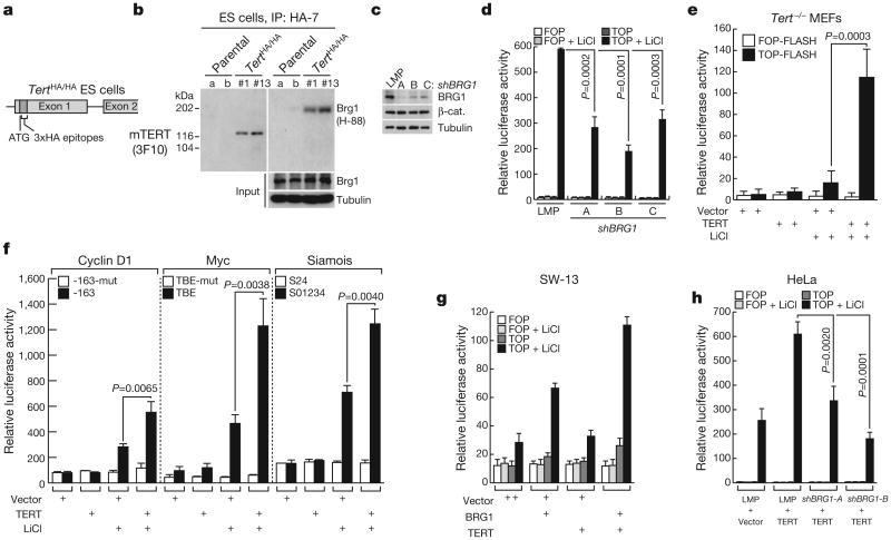
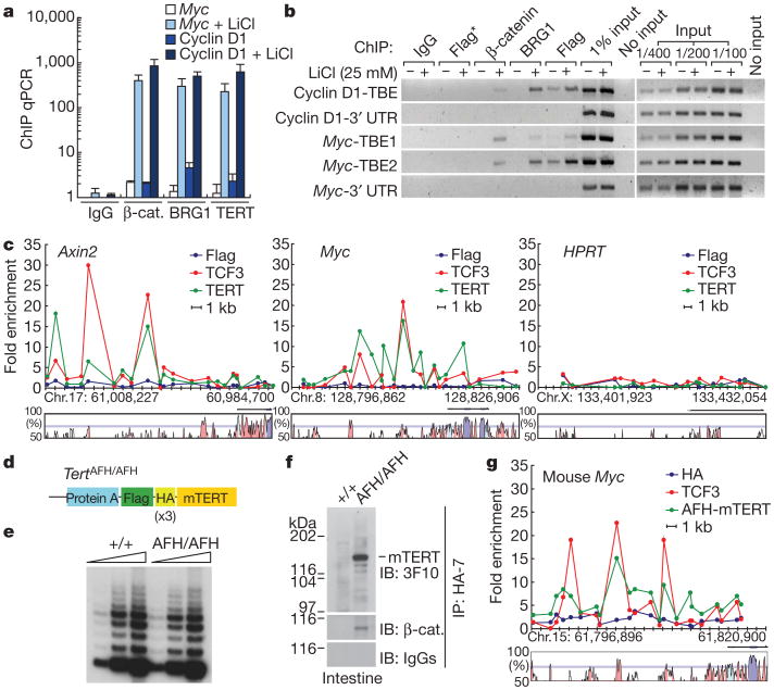
Stem cells are controlled, in part, by genetic pathways frequently dysregulated during human tumorigenesis. Either stimulation of Wnt/beta-catenin signalling or overexpression of telomerase is sufficient to activate quiescent epidermal stem cells in vivo, although the mechanisms by which telomerase exerts these effects are not understood. Here we show that telomerase directly modulates Wnt/beta-catenin signalling by serving as a cofactor in a beta-catenin transcriptional complex. The telomerase protein component TERT (telomerase reverse transcriptase) interacts with BRG1 (also called SMARCA4), a SWI/SNF-related chromatin remodelling protein, and activates Wnt-dependent reporters in cultured cells and in vivo. TERT serves an essential role in formation of the anterior-posterior axis in Xenopus laevis embryos, and this defect in Wnt signalling manifests as homeotic transformations in the vertebrae of Tert(-/-) mice. Chromatin immunoprecipitation of the endogenous TERT protein from mouse gastrointestinal tract shows that TERT physically occupies gene promoters of Wnt-dependent genes. These data reveal an unanticipated role for telomerase as a transcriptional modulator of the Wnt/beta-catenin signalling pathway.


Figures

Figure 1
Figure 1. TERT activates Wnt reporter plasmids in a BRG1-dependent manner
a, Diagram of HA–TERT knock-in ES cells. b, Interaction of endogenous TERT with BRG1 in TertHA/HA ES cells by immunoprecipitation (IP) and immunoblot (IB). HA-7 and 3F10, anti-HA antibodies. c, Depletion of BRG1 protein by shRNAs in HeLa cells by immunoblot. d, TOP-FLASH reporter activity in HeLa cells transduced with empty vector (LMP) or shRNA BRG1 retroviruses, then transfected with TOP-FLASH plasmid (wild-type TCF sites) and treated with LiCl (n = 3) (FOP-FLASH, mutant TCF sites). e, TOP-FLASH activity in Tert−/− MEFs co-transfected with empty vector or mouse TERT expression plasmid and treated with or without LiCl (n = 4). f, Luciferase activity after transient co-transfection of reporter plasmids comprising cyclin D1, Myc, or siamois promoters driving luciferase with TERT plasmid or empty vector in HeLa cells, followed by LiCl treatment (n = 3). Filled bar, wild-type TCF binding elements (TBE); open bar, mutant TBEs. g, Effect on TOP-FLASH activity of transient co-transfection of BRG1, TERT or BRG1 combined with TERT in SW-13 cells lacking BRG1 (n = 2). h, Effect of BRG1 depletion with shRNA on TERT-mediated activation of TOP-FLASH activity in HeLa cells (n = 3). Error bars indicate standard deviation; P values produced by Student's t-test.
Figure 2
Figure 2. TERT activates the Wnt pathway in vivo and is required for efficient target gene activation by WNT3A ligand in mouse ES cells
a, b, X-Gal staining for β-galactosidase activity in small intestine and colon of Axin2lacZ/+ reporter mice, i-Tertci Axin2lacZ/+ mice or controls. Whole mounts seen from abluminal side (a); histology, nuclear fast red (b). Arrowheads indicate crypts. Doxy, doxycycline; s.i., small intestine. c, CD44 protein in small intestine crypts by immunofluorescence. d, Schematic for deletion of TERT by 4-OHT treatment of conditional TERT knockout (CKO) ES cells. e, Cre-mediated recombination in ES cells by Southern blot. Arrowhead, recombined Tert allele. f, Induction of Axin2 by WNT3A ligand in TERT conditional knockout (CKO) mouse ES cells treated with vehicle or with 250nM 4-OHT for 3 days, exposed to WNT3A (100 ng ml−1) for 24 h and analysed by qPCR (n = 3). g, h, Basal expression of Axin2 mRNA by qPCR in TERT conditional knockout mouse ES cells treated with vehicle or 4-OHT, and Axin2 mRNA levels in TERT conditional knockout cells with stable overexpression of mouse TERTci (n = 3), shown in h by immunoprecipitation and western blot analysis. Error bars indicate s.d. Original magnification: a, ×4 (insets ×8); b, ×20; c, ×40.
Figure 3
Figure 3. TERT promotes anterior–posterior axis duplication and is required for efficient anterior–posterior axis in Xenopus
a, b, Duplicate anterior–posterior axis formation in Xenopus embryos co-injected with Xenopus(x)β-catenin mRNA (0.2 ng) and increasing amounts of xTert mRNA. Open arrowheads, extra axes. c, Duplicate axis with co-injection of xTertci and xβ-catenin mRNA (0.4 ng). d, TRAP activity at blastula, gastrula and neurula stages in embryos injected with TERT morpholino (TMO2) or control (StdMO). e, Defects in anterior–posterior axis development in embryos injected with TERT morpholinos (TMO1 or TMO2), but not StdMO, scored using dorso-anterior index (DAI). f, Ectopic neural tube formation (arrowhead) in TMO2-injected embryos. ar, archenteron; dnt, dorsal neural tube; en, endoderm; ep, epidermis; nc, notochord; vnt, ventral neural tube. Transverse sections; haematoxylin and eosin staining. g, Rescue of developmental phenotypes with xTERT, xTERTci and human (h)TERTci, stages 37–38. h, TOP-FLASH analysis by co-injecting reporter plasmid at the 2-cell stage with either StdMO or TMO1, or co-injecting reporter plasmid with β-gal, xTERT or β-catenin (n = 3, P values produced by Student's t-test, error bars indicate s.d.).
Figure 4
Figure 4. Somite defects in Xenopus embryos treated with TERT morpholino and homeotic transformations in Tert−/− mice
a, Somite staining using 12/101 antibody of Xenopus embryos injected with either StdMO or TMO2. Arrows show somite boundaries. b, WNT3A-mediated induction of Cdx1 in TERT conditional knockout ES cells treated with vehicle or 4-OHT for 3 days, followed by WNT3A for 24 h. * P = 0.002 by Student's t-test (n = 3). c, Levels of xCdx1, xCdx2and xCdx4measured by qPCR in Xenopus embryos injected with StdMO or TMO2 and collected at the late gastrulation stage. *P = 0.0066, **P = 0.0003, ***P < 0.0001 by Student's t-test (n = 4). d, e, T13 to L1 vertebral homeotic transformations in G1 Tert−/− mice. Unilateral loss of 13th rib (middle) or bilateral loss of 13th ribs (right). Arrowhead, 13th rib remnant; arrows, missing 13th ribs. Adult skeletons were stained with alcian blue and alizarin red (ventral; d). Three-dimensional reconstruction of microCT scan images of independent mice (dorsal; e).f, Summary of axial skeletal defects in G1 Tert−/− mice. P = 0.0046 by Fisher's exact test.
Figure 5
Figure 5. TERT occupies Wnt target gene promoters in HeLa cells and in mouse small intestine
a, b, Association of Flag–TERT, BRG1 or β-catenin with TBE-containing fragments of the cyclin D1 and Myc promoters in Flag–TERT HeLa cells with or without LiCl treatment by ChIP qPCR (a) and semi-qPCR (b). Error bars indicate s.d. 3′ UTR sequences lacking TBEs, negative controls. Flag*, Flag ChIP from HeLa parental cells. c, Scanning ChIP across 20 kb of Axin2, Myc and HPRT promoters in lithium-treated HeLa parental and Flag–TERT HeLa cells by qPCR. Non-coding sequences conserved between human and mouse genomes are shown (VISTA graphs: exon, purple; 5′ UTR, blue; conserved non-coding sequence, pink). d, AFH-TERT knock-in mouse allele. e, Telomerase activity in small intestine from wild-type and TertAFH/AFH mice (TRAP). f, Association of AFH-TERT and β-catenin at the endogenous level in extracts from small intestine of TertAFH/AFH mice. Anti-HA antibody immunoprecipitation, followed by immunoblot with anti-HA antibody or anti-b-catenin antibody. IgGs, negative control for immunoblot. g, In vivo ChIP from Tert+/+ and TertAFH/AFH mouse small intestine shows co-occupancy across 20 kb of the Myc promoter for TCF3 (red) and AFH-TERT (green). ChIP from Tert+/+ mouse with anti-HA-7 antibody, negative control (blue).

Comment in

References

    1. Reya T, Clevers H. Wnt signalling in stem cells and cancer. Nature. 2005;434:843–850. - PubMed
    1. Gat U, DasGupta R, Degenstein L, Fuchs E. De Novo hair follicle morphogenesis and hair tumors in mice expressing a truncated β-catenin in skin. Cell. 1998;95:605–614. - PubMed
    1. Van Mater D, Kolligs FT, Dlugosz AA, Fearon ER. Transient activation of β-catenin signaling in cutaneous keratinocytes is sufficient to trigger the active growth phase of the hair cycle in mice. Genes Dev. 2003;17:1219–1224. - PMC - PubMed
    1. Lo Celso C, Prowse DM, Watt FM. Transient activation of β-catenin signalling in adult mouse epidermis is sufficient to induce new hair follicles but continuous activation is required to maintain hair follicle tumours. Development. 2004;131:1787–1799. - PubMed
    1. Sarin KY, et al. Conditional telomerase induction causes proliferation of hair follicle stem cells. Nature. 2005;436:1048–1052. - PMC - PubMed

Publication types

MeSH terms