Acquisition of cell-cell fusion activity by amino acid substitutions in spike protein determines the infectivity of a coronavirus in cultured cells
- PMID: 19572016
- PMCID: PMC2700284
- DOI: 10.1371/journal.pone.0006130
Acquisition of cell-cell fusion activity by amino acid substitutions in spike protein determines the infectivity of a coronavirus in cultured cells
Abstract
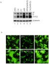
Coronavirus host and cell specificities are determined by specific interactions between the viral spike (S) protein and host cell receptor(s). Avian coronavirus infectious bronchitis (IBV) has been adapted to embryonated chicken eggs, primary chicken kidney (CK) cells, monkey kidney cell line Vero, and other human and animal cells. Here we report that acquisition of the cell-cell fusion activity by amino acid mutations in the S protein determines the infectivity of IBV in cultured cells. Expression of S protein derived from Vero- and CK-adapted strains showed efficient induction of membrane fusion. However, expression of S protein cloned from the third passage of IBV in chicken embryo (EP3) did not show apparent syncytia formation. By construction of chimeric S constructs and site-directed mutagenesis, a point mutation (L857-F) at amino acid position 857 in the heptad repeat 1 region of S protein was shown to be responsible for its acquisition of the cell-cell fusion activity. Furthermore, a G405-D point mutation in the S1 domain, which was acquired during further propagation of Vero-adapted IBV in Vero cells, could enhance the cell-cell fusion activity of the protein. Re-introduction of L857 back to the S gene of Vero-adapted IBV allowed recovery of variants that contain the introduced L857. However, compensatory mutations in S1 and some distant regions of S2 were required for restoration of the cell-cell fusion activity of S protein carrying L857 and for the infectivity of the recovered variants in cultured cells. This study demonstrates that acquisition of the cell-cell fusion activity in S protein determines the selection and/or adaptation of a coronavirus from chicken embryo to cultured cells of human and animal origins.
Conflict of interest statement
Figures


Similar articles
-
Roles in cell-to-cell fusion of two conserved hydrophobic regions in the murine coronavirus spike protein.Virology. 1998 May 10;244(2):483-94. doi: 10.1006/viro.1998.9121. Virology. 1998. PMID: 9601516 Free PMC article.
-
Proteolytic activation of the spike protein at a novel RRRR/S motif is implicated in furin-dependent entry, syncytium formation, and infectivity of coronavirus infectious bronchitis virus in cultured cells.J Virol. 2009 Sep;83(17):8744-58. doi: 10.1128/JVI.00613-09. Epub 2009 Jun 24. J Virol. 2009. PMID: 19553314 Free PMC article.
-
Genetic analysis of the SARS-coronavirus spike glycoprotein functional domains involved in cell-surface expression and cell-to-cell fusion.Virology. 2005 Oct 25;341(2):215-30. doi: 10.1016/j.virol.2005.06.046. Epub 2005 Aug 15. Virology. 2005. PMID: 16099010 Free PMC article.
-
Emergence of a coronavirus infectious bronchitis virus mutant with a truncated 3b gene: functional characterization of the 3b protein in pathogenesis and replication.Virology. 2003 Jun 20;311(1):16-27. doi: 10.1016/s0042-6822(03)00117-x. Virology. 2003. PMID: 12832199 Free PMC article.
-
Site directed mutagenesis of the murine coronavirus spike protein. Effects on fusion.Adv Exp Med Biol. 1995;380:283-6. doi: 10.1007/978-1-4615-1899-0_45. Adv Exp Med Biol. 1995. PMID: 8830493 Review.
Cited by
-
Contributions of the S2 spike ectodomain to attachment and host range of infectious bronchitis virus.Virus Res. 2013 Nov 6;177(2):127-37. doi: 10.1016/j.virusres.2013.09.006. Epub 2013 Sep 13. Virus Res. 2013. PMID: 24041648 Free PMC article.
-
Regulation of the ER Stress Response by the Ion Channel Activity of the Infectious Bronchitis Coronavirus Envelope Protein Modulates Virion Release, Apoptosis, Viral Fitness, and Pathogenesis.Front Microbiol. 2020 Jan 24;10:3022. doi: 10.3389/fmicb.2019.03022. eCollection 2019. Front Microbiol. 2020. PMID: 32038520 Free PMC article.
-
Binding of the 5'-untranslated region of coronavirus RNA to zinc finger CCHC-type and RNA-binding motif 1 enhances viral replication and transcription.Nucleic Acids Res. 2012 Jun;40(11):5065-77. doi: 10.1093/nar/gks165. Epub 2012 Feb 22. Nucleic Acids Res. 2012. PMID: 22362731 Free PMC article.
-
Molecular Evolution of Human Coronavirus Genomes.Trends Microbiol. 2017 Jan;25(1):35-48. doi: 10.1016/j.tim.2016.09.001. Epub 2016 Oct 19. Trends Microbiol. 2017. PMID: 27743750 Free PMC article. Review.
-
The interactions of folate with the enzyme furin: a computational study.RSC Adv. 2021 Jul 6;11(38):23815-23824. doi: 10.1039/d1ra03299b. eCollection 2021 Jul 1. RSC Adv. 2021. PMID: 35479793 Free PMC article.
References
-
- Wege H, Siddell S, Ter Meulen V. The biology and pathogenesis of coronavirus. Curr Top Microbiol Immunol. 1982;99:165–200. - PubMed
-
- Guan Y, Zheng BJ, He YQ, Liu XL, Zhuang ZX, et al. Isolation and characterization of viruses related to the SARS coronavirus from animals in southern China. Science. 2003;302:276–278. - PubMed
-
- Li W, Shi Z, Yu M, Ren W, Smith C, et al. Bats are natural reservoirs of SARS-like coronaviruses. Science. 2005;310:676–979. - PubMed
Publication types
MeSH terms
Substances
LinkOut - more resources
Full Text Sources
Other Literature Sources
Research Materials

