

Sihler's whole mount nerve staining technique: a review
- PMID: 19572223
- PMCID: PMC2822894
- DOI: 10.3109/10520290903048384
Sihler's whole mount nerve staining technique: a review
Abstract
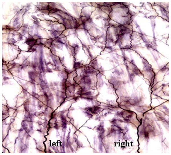
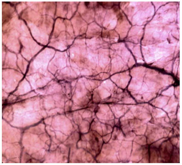
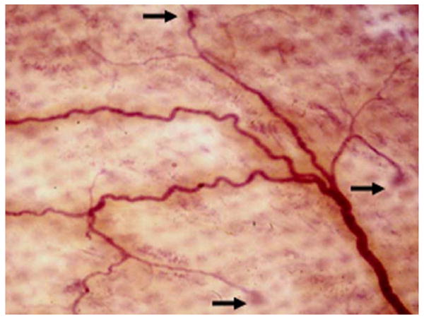
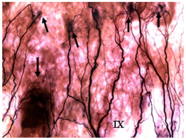
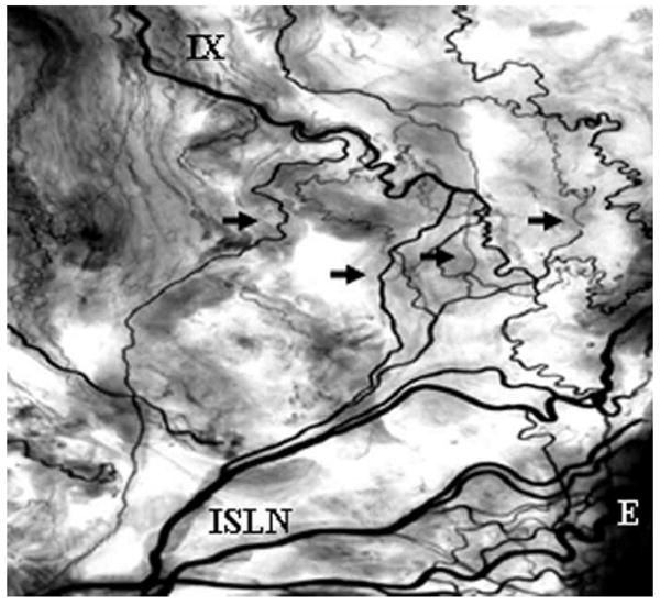
Sihler's stain is a whole mount nerve staining technique that renders other soft tissue translucent or transparent while staining the nerves. It permits mapping of entire nerve supply patterns of organs, skeletal muscles, mucosa, skin, and other structures after the specimens are fixed in neutralized formalin, macerated in potassium hydroxide, decalcified in acetic acid, stained in Ehrlich's hematoxylin, destained in acetic acid, and cleared in glycerin. The unique advantage of Sihler's stain over other anatomical methods is that all the nerves within the stained specimen can be visualized in their three-dimensional positions. To date, Sihler's stain is the best tool for demonstrating the precise intramuscular branching and distribution patterns of skeletal muscles, which are important not only for anatomists, but also for physiologists and clinicians. Advanced knowledge of the neural structures within mammalian skeletal muscles is critical for understanding muscle functions, performing electrophysiological experiments and developing novel neurosurgical techniques. In this review, Sihler's stain is described in detail and its use in nerve mapping is surveyed. Special emphasis is placed on staining procedures and troubleshooting, strengths and limitations, applications, major contributions to neuroscience, physiological and clinical significance, and areas for further technical improvement that deserve future research.
Conflict of interest statement
Figures

Similar articles
-
[Application of the modified Sihler's stain technique to cadaveric peripheral nerves after medical students' dissection course].Kaibogaku Zasshi. 2005 Sep;80(3):67-72. Kaibogaku Zasshi. 2005. PMID: 16196427 Japanese.
-
Sihler's staining technique: How to and guidance for botulinum neurotoxin injection in human muscles.Clin Anat. 2024 Mar;37(2):169-177. doi: 10.1002/ca.24076. Epub 2023 May 31. Clin Anat. 2024. PMID: 37255275 Review.
-
Clinical and anatomical approach using Sihler's staining technique (whole mount nerve stain).Anat Cell Biol. 2011 Mar;44(1):1-7. doi: 10.5115/acb.2011.44.1.1. Epub 2011 Mar 31. Anat Cell Biol. 2011. PMID: 21519543 Free PMC article.
-
Modified Sihler's technique for studying the distribution of intramuscular nerve branches in mammalian skeletal muscle.Anat Rec. 1997 Jan;247(1):137-44. doi: 10.1002/(SICI)1097-0185(199701)247:1<137::AID-AR16>3.0.CO;2-Q. Anat Rec. 1997. PMID: 8986311
-
Guidance in botulinum neurotoxin injection for lower extremity spasticity: Sihler's staining technique.Surg Radiol Anat. 2023 Aug;45(8):1055-1062. doi: 10.1007/s00276-023-03178-9. Epub 2023 Jun 9. Surg Radiol Anat. 2023. PMID: 37294437 Review.
Cited by
-
An unusual variant of the abducens nerve duplication with two nerve trunks merging within the orbit: a case report with comments on developmental background.Surg Radiol Anat. 2016 Jul;38(5):625-9. doi: 10.1007/s00276-015-1573-x. Epub 2015 Oct 26. Surg Radiol Anat. 2016. PMID: 26501961 Free PMC article.
-
Comparison of lateral and medial rectus muscle in human: an anatomical study with particular emphasis on morphology, intramuscular innervation pattern variations and discussion on clinical significance.Surg Radiol Anat. 2020 May;42(5):607-616. doi: 10.1007/s00276-019-02400-x. Epub 2020 Jan 2. Surg Radiol Anat. 2020. PMID: 31897658
-
Activation of peripheral nerve fibers by electrical stimulation in the sole of the foot.BMC Neurosci. 2013 Oct 8;14:116. doi: 10.1186/1471-2202-14-116. BMC Neurosci. 2013. PMID: 24103294 Free PMC article.
-
Reinnervation of denervated muscle by implantation of nerve-muscle-endplate band graft to the native motor zone of the target muscle.Brain Behav. 2017 May 3;7(6):e00668. doi: 10.1002/brb3.668. eCollection 2017 Jun. Brain Behav. 2017. PMID: 28638701 Free PMC article.
-
Nerve-muscle-endplate band grafting: a new technique for muscle reinnervation.Neurosurgery. 2011 Dec;69(2 Suppl Operative):ons208-24; discussion ons224. doi: 10.1227/NEU.0b013e31822ed596. Neurosurgery. 2011. PMID: 21796004 Free PMC article.
References
-
- Amirali A, Mu L, Sunkara M, Gracies JM, Simpson DM. Anatomical localization of motor endplate bands in the human biceps brachii. J Clin Neuromusc Dis. 2007;9:306–312. - PubMed
-
- Bae YC, Zuker RM, Manktelow RT, Wade S. A comparison of commissure excursion following gracilis muscle transplantation for facial paralysis using a crossface nerve graft versus the motor nerve to the masseter nerve. Plast Reconstr Surg. 2006;117:2407–2413. - PubMed
-
- Bayramicli M, Jackson IT, Herschman B. Innervation of skin grafts over free muscle flaps. Br J Plast Surg. 2000;53:130–136. - PubMed
-
- Berkowitz RG, Sun QJ, Chalmers J, Pilowsky PM. Respiratory activity of the rat posterior cricoarytenoid muscle. Ann Otol Rhinol Laryngol. 1997;106:897–901. - PubMed
-
- Bilge O, Pinar Y, Ozer MA, Govsa F. The vascular anatomy of the lumbrical muscles in the hand. J Plast Reconstr Aesthet Surg. 2007;60:1120–1126. - PubMed
Publication types
MeSH terms
Substances
Grants and funding
LinkOut - more resources
Full Text Sources