. 2009 Jul 7;16(1):33-43.
doi: 10.1016/j.ccr.2009.05.008.

Increased radioresistance and accelerated B cell lymphomas in mice with Mdmx mutations that prevent modifications by DNA-damage-activated kinases

Affiliations

Increased radioresistance and accelerated B cell lymphomas in mice with Mdmx mutations that prevent modifications by DNA-damage-activated kinases

Yunyuan V Wang et al. Cancer Cell. .

Abstract

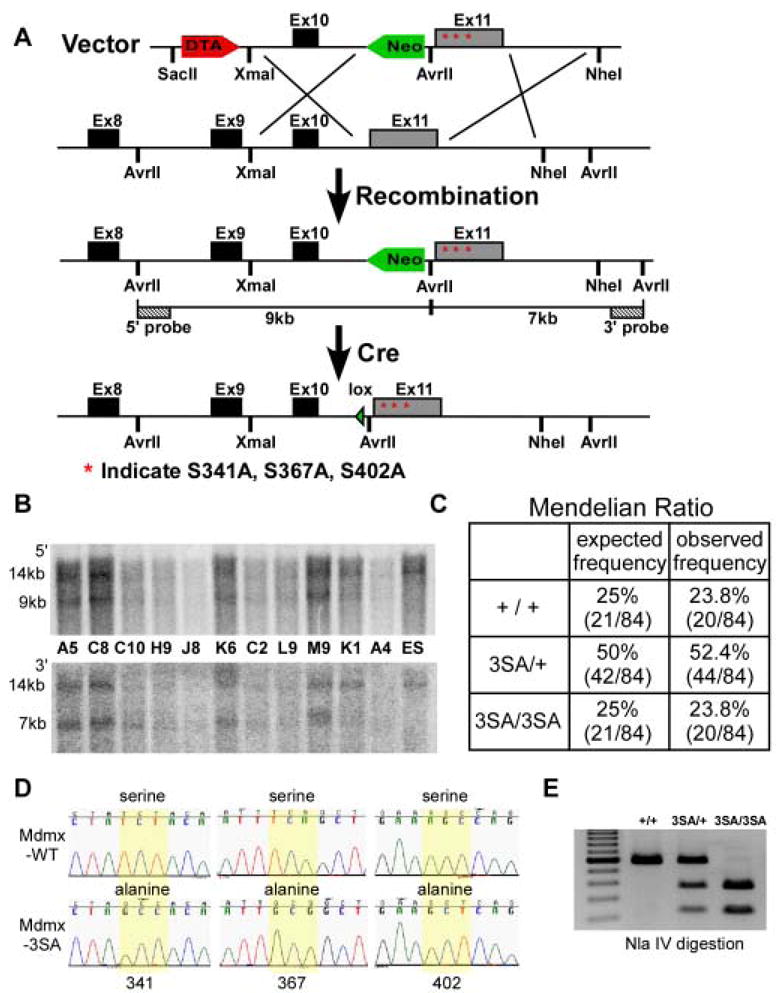
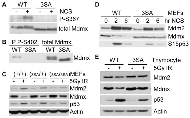
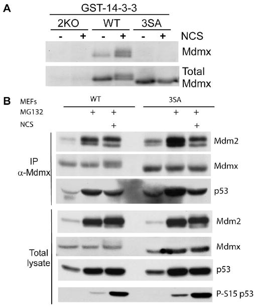
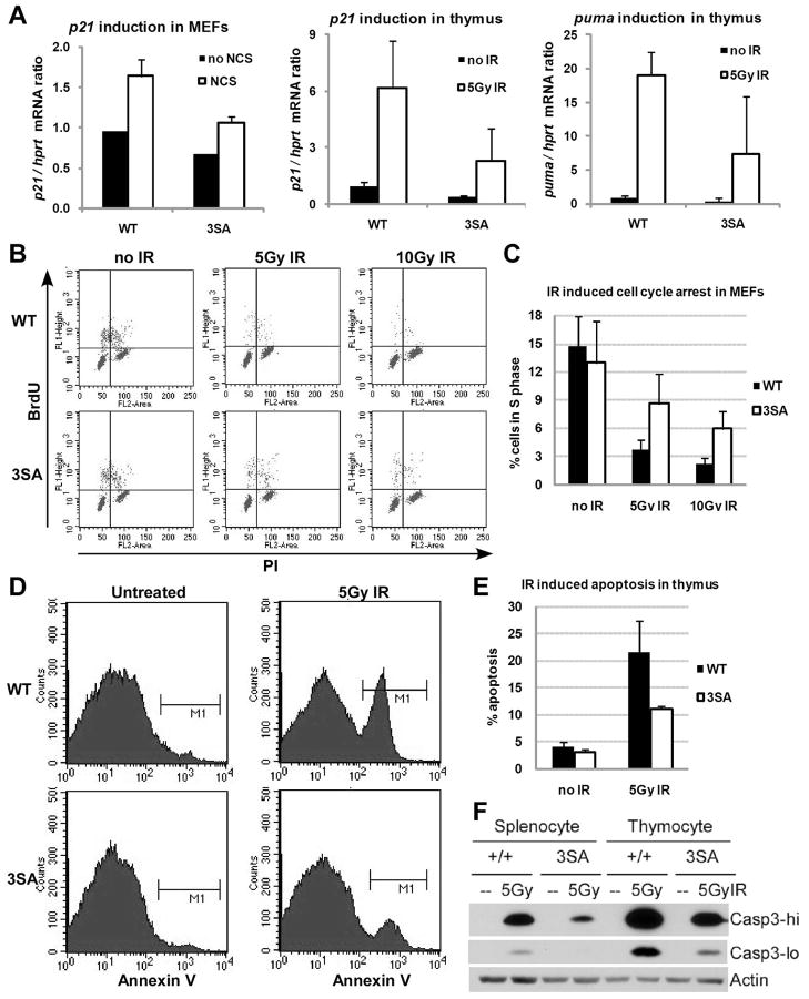
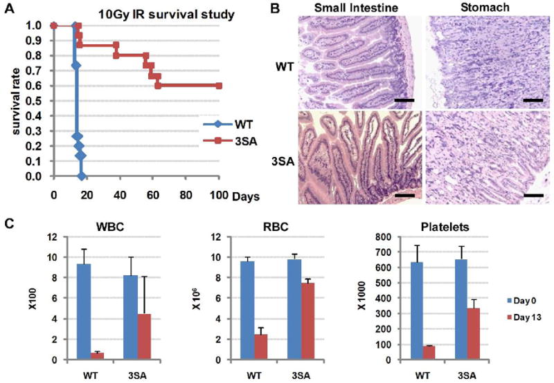
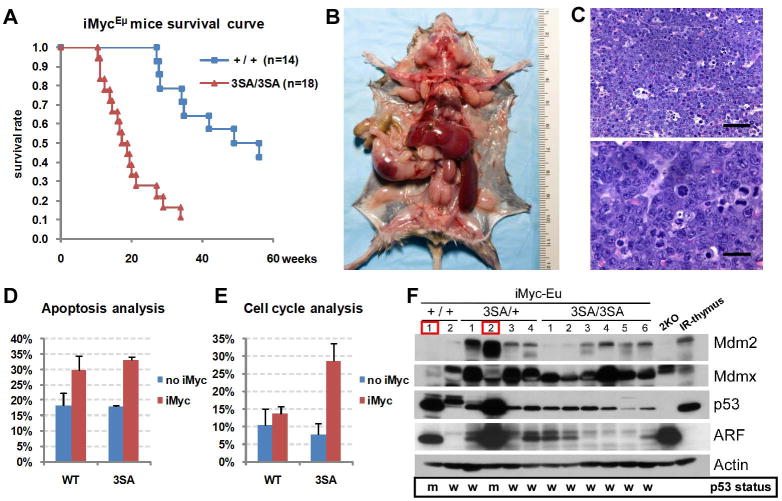
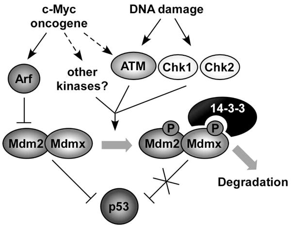
Mdmx is a critical negative regulator of the p53 pathway that is stoichiometrically limiting in some tissues. Posttranslational modification and degradation of Mdmx after DNA damage have been proposed to be essential for p53 activation. We tested this model in vivo, where critical stoichiometric relationships are preserved. We generated an Mdmx mutant mouse in which three conserved serines (S341, S367, S402) targeted by DNA-damage-activated kinases were replaced by alanines to investigate whether modifications of these residues are important for Mdmx degradation and p53 activation. The mutant mice were remarkably resistant to radiation, and very susceptible to Myc-induced lymphomagenesis. These data demonstrate that Mdmx downregulation is crucial for effective p53-mediated radiation responses and tumor suppression in vivo.


Figures

Figure 1
Figure 1. Generation of Mdmx3SA allele and genotype analysis of Mdmx3SA
(A) Schematic representation of the 3′ end of the mouse Mdmx locus, the targeting vector and homologous recombined Mdmx3SA allele, followed by excision of loxP-flanked Neo cassette. The targeting construct contains a DTA negative selection marker, 5′ homology region with exon10, a loxP flanked Neo positive selection cassette, followed by the exon11 with Mdmx mutations indicated as stars and 5′ homology region. The insertion of Neo introduces an additional Avr II site that was used for Southern analysis with the indicated external 5′ and 3′ probes. Cre expression in the male germline subsequently allows excision of Neo. (B) Southern blot analysis of targeted ES clones. The 14kb band represents the Avr II fragments from the wild type allele using both 5′ and 3′ probes. The recombined allele generates a 9kb fragment detected by the 5′ probe and a 7kb fragment detected by the 3′ probe. (C) The expected and observed Mendelian ratios from Mdmx3SA heterozygote breeding. (D) Sequences of Mdmx transcript expressed from MdmxWT and Mdmx3SA thymus. Yellow highlight shows that the codons that encode S341, S367 and S402 of the wild type Mdmx are mutated in the Mdmx3SA allele. (E) PCR Genotype of the Mdmx3SA mutant. PCR amplifies the region surrounding the mutations to produce a 579bp fragment. The introduction of mutations generates an Nla IV site that results in 366bp and 213bp fragments when PCR product is digested with Nla IV.
Figure 2
Figure 2. Mdmx3SA mutations impair DNA damage-induced Mdmx degradation
(A) The MdmxWT and Mdmx3SA MEFs were pre-incubated with MG132 (10 μM) for 30 minutes, followed by 2 hrs of NCS (200 ng/ml) incubation. Lysates were immunoprecipitated with anti-Mdmx antibody, and blotted with anti-phospho-S367 antibody. (B) MEFs were treated as described in (A). Lysates were immunoprecipitated with anti-phospho-S402 Mdmx antibody, and blotted with anti-Mdmx antibody. (C) MEFs were irradiated with 5Gy of γ-irradiation. Four hours later, cells were lysed for western analysis. (D) MEFs were treated with 200ng/ml of NCS for 0, 2 and 6 hrs. Lysates were analyzed by western blotting using antibodies against Mdm2, Mdmx and phospho-S15 of p53. (E) Thymocytes were irradiated with 5Gy of γ-irradiation. Four hours later, cells were lysed for western analysis.
Figure 3
Figure 3. Analyses of protein interactions with Mdmx3SA in response to DNA damage
(A) Cell lysates from MdmxWT, Mdmx3SA, 2KO (p53−/−;Mdmx−/−) MEFs either untreated or treated with NCS (200 ng/ml) in the presence of MG132 (10 μM) for 2 hrs were incubated with glutathione beads pre-bound with GST-14-3-3ε proteins. Bead-bound proteins were eluted and analyzed by western blotting. (B) MEFs were either untreated or treated with MG132 (10 μM) or treated with MG132 plus NCS (200 ng/ml) and lysed 2hrs after. Cell lysates were immunoprecipitated with anti-Mdmx antibody, blotted with anti-Mdm2, anti-Mdmx and anti-p53 antibodies. Anti-phospho-S15 of p53 antibody was used as a control for detecting the DNA damage signal induced by NCS.
Figure 4
Figure 4. p53 activity is reduced in Mdmx3SA mutant
(A) MEFs were treated with 500ng/ml of NCS or 5 week-old mice were exposed to 5Gy of γ-irradiation. Six hours after NCS incubation or 4 hrs after irradiation, cells or thymus removed from MdmxWT (WT) and Mdmx3SA (3SA) mice were lysed for RNA isolation. p21 and puma gene expression was analyzed by RT-qPCR. Results were normalized to hprt mRNA. (B) Cell cycle arrest analysis. MEFs were either untreated or exposed to 5Gy or 10Gy of γ-irradiation. Twenty-three hours after irradiation, 10μM of BrdU was added in the cell culture for one hour before FACS analysis. BrdU-positive cells that represent cells in S-phase during BrdU incorporation were quantified in (C). Error bars represent +/−SD from three independent experiments. (D) Analysis of apoptosis in vivo. Mice were untreated or exposed to 5Gy of γ-irradiation. Four hours later thymocytes were isolated from mice and stained with Annexin V-FITC for FACS analysis. Annexin V positive cells that represent apoptotic cells were quantified in (E). Error bars represent +/−SD from three independent experiments. (F) Apoptosis in splenocytes and thymocytes. Mice were treated as described in (D). Splenocytes and thymocytes were isolated and analyzed for the expression of active form of Caspase-3 by western blotting with high exposure (hi) or low exposure (lo).
Figure 5
Figure 5. Mdmx3SA mice are resistant to a lethal dose of ionizing irradiation
(A) Kaplan-Meier survival curves of fifteen age-matched MdmxWT (WT, n=15) and Mdmx3SA homozygote (3SA, n=15) mice following exposure to 10Gy of whole-body γ-irradiation. (B) Histology of the gastrointestinal tract from MdmxWT and Mdmx3SA mice showing no lesions associated with irradiation (H&E, Bar = 100 μm). (C) Complete blood count analysis of white blood cells (WBC), red blood cells (RBC) and platelets from MdmxWT and Mdmx3SA mice, 13 days post 10Gy of whole-body γ-irradiation. Error bars are +/−SD from 3 animals. μ
Figure 6
Figure 6. Myc-induced tumorigenesis is accelerated in Mdmx3SA mice
(A) Kaplan-Meier survival curves of iMyc;+/+ (n=14) and iMyc;3SA/3SA (n=18) mice. (B) Early onset lymphoma in a 113 day-old iMyc;3SA/3SA mouse: dissection reveals severe enlargement of the spleen, liver and most lymph nodes (notably, the mesenteric, cervical, brachial, axillary, mediastinal, pancreatic, renal, inguinal and lumbar nodes). (C) High grade B-cell lymphoma in iMyc;3SA/3SA mice. Upper panel: the lymph node architecture is replaced with solid sheets of intermediate size lymphocytes admixed with apoptotic tumor cells and large macrophages containing tingible bodies producing a typical “starry-sky” effect (H&E, Bar = 50 μm). Lower panel: Intermediate size lymphocytes with moderate to marked anisocytosis/anisokaryosis, high mitotic index, abnormal mitosis and apoptotic tumor cells (H&E, Bar = 20 μm). (D) Apoptosis analysis. Splenocytes were isolated from 8 week-old littermate mice and stained with Annexin V-FITC for FACS analysis. Annexin V positive cells were quantified. Error bars represent +/−SD from analysis. Eight week-old littermate mice that contain were i.p. injected with 100mg/kg of BrdU. Five hours later, and stained with anti-BrdU antibody for FACS analysis represent cells in S-phase were quantified. Error bars animals. (F) Levels of Mdm2, Mdmx, p53, Arf assessed extracts from iMyc;+/+, iMyc;3SA/+ and iMyc;3SA/3SA mice. p53−/− Mdm2−/− MEFs(2KO) and thymus irradiated in vivo (IR-thymus) were panel shows the p53 status in those tumors (w indicates mutated p53). Tumor 1 from iMyc;+/+ and Tumor 2 from red contain p53 mutations at R172H (CGC → CAC) and K129T (AAG → ACG) respectively (see Figure S8 for details).
Figure 7
Figure 7. Mechanism for the role of Mdmx modification in DNA damage and oncogenic signaling pathway
Phosphorylations of Mdmx can be initiated by either DNA damage signaling or by activated oncogenes. Phosphorylations of Mdmx modulate its interaction with other proteins, such as 14-3-3, the protein analyzed in this study. Interaction of Mdmx and 14-3-3 is required for accelerated Mdmx and Mdm2 degradation after DNA damage, probably by limiting Mdm2/Mdmx interaction with the deubiquitylase HAUSP (see text). Preferential degradation of Mdm2 and Mdmx reduces their ability to interact with and antagonize p53 function, leading to p53 activation. The 3SA mutations introduced in this study prevent 14-3-3 interaction after DNA damage, leading to Mdmx-Mdm2 stabilization, and a corresponding increased ability to degrade p53.

Comment in

  • Tumour suppression: Full on.
    Alderton GK. Alderton GK. Nat Rev Cancer. 2009 Sep;9(9):612. doi: 10.1038/nrc2725. Nat Rev Cancer. 2009. PMID: 19705529 No abstract available.

References

    1. Adams J, Harris A, Pinkert C, Corcoran L, Alexander W, Cory S, Palmiter R, Brinster R. The c-myc oncogene driven by immunoglobulin enhancers induces lymphoid malignancy in transgenic mice. Nature. 1985;318(6046):533–8. - PubMed
    1. Alitalo K, Koskinen P, Mäkelä TP, Saksela K, Sistonen L, Winqvist R. myc oncogenes: activation and amplification. Biochimica et Biophysica Acta (BBA) - Reviews on Cancer. 1987;907(1):1. - PubMed
    1. Bartkova J, Horejsi Z, Koed K, Kramer A, Tort F, Zieger K, Guldberg P, Sehested M, Nesland JM, Lukas C, et al. DNA damage response as a candidate anti-cancer barrier in early human tumorigenesis. Nature. 2005;434(7035):864. - PubMed
    1. Burdelya LG, Komarova EA, Hill JE, Browder T, Tararova ND, Mavrakis L, DiCorleto PE, Folkman J, Gudkov AV. Inhibition of p53 Response in Tumor Stroma Improves Efficacy of Anticancer Treatment by Increasing Antiangiogenic Effects of Chemotherapy and Radiotherapy in Mice. Cancer Res. 2006;66(19):9356–9361. - PubMed
    1. Chen L, Gilkes DM, Pan Y, Lane WS, Chen J. ATM and Chk2-dependent phosphorylation of MDMX contribute to p53 activation after DNA damage. Embo J. 2005;24(19):3411–22. - PMC - PubMed

Publication types

MeSH terms