. 2009 Sep 11;284(37):24806-15.
doi: 10.1074/jbc.M109.018424. Epub 2009 Jul 2.

Evidence for catalytic roles for Plasmodium falciparum aminopeptidase P in the food vacuole and cytosol

Affiliations

Evidence for catalytic roles for Plasmodium falciparum aminopeptidase P in the food vacuole and cytosol

Daniel Ragheb et al. J Biol Chem. .

Abstract

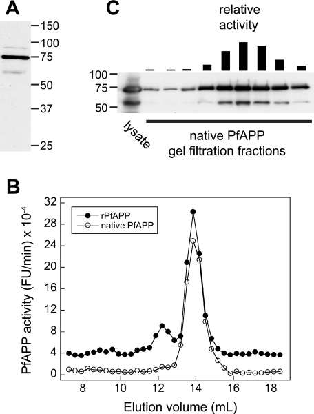
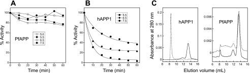
The metalloenzyme aminopeptidase P catalyzes the hydrolysis of amino acids from the amino termini of peptides with a prolyl residue in the second position. The human malaria parasite Plasmodium falciparum expresses a homolog of aminopeptidase P during its asexual intraerythrocytic cycle. P. falciparum aminopeptidase P (PfAPP) shares with mammalian cytosolic aminopeptidase P a three-domain, homodimeric organization and is most active with Mn(II) as the cofactor. A distinguishing feature of PfAPP is a 120-amino acid amino-terminal extension that appears to be removed from the mature protein. PfAPP is present in the food vacuole and cytosol of the parasite, a distribution that suggests roles in vacuolar hemoglobin catabolism and cytosolic peptide turnover. To evaluate the plausibility of these putative functions, the stability and kinetic properties of recombinant PfAPP were evaluated at the acidic pH of the food vacuole and at the near-neutral pH of the cytosol. PfAPP exhibited high stability at 37 degrees C in the pH range 5.0-7.5. In contrast, recombinant human cytosolic APP1 was unstable and formed a high molecular weight aggregate at acidic pH. At both acidic and slightly basic pH values, PfAPP efficiently hydrolyzed the amino-terminal X-Pro bond of the nonapeptide bradykinin and of two globin pentapeptides that are potential in vivo substrates. These results provide support for roles for PfAPP in peptide catabolism in both the food vacuole and the cytosol and suggest that PfAPP has evolved a dual distribution in response to the metabolic needs of the intraerythrocytic parasite.


Figures

FIGURE 1.
FIGURE 1.
Localization of native PfAPP. A, indirect immunofluorescence localization of PfAPP in aldehyde-fixed trophozoite-stage parasites. Left column, anti-PfAPP-associated fluorescence; middle column, phase contrast image; right column, relative fluorescence intensity along a line bisecting the parasite. The food vacuole is indicated by an arrowhead. Bar, 2 μm. B, localization of PfAPP by cryo-immunoelectron microscopy. rbc, red blood cell; fv, food vacuole; pvm/ppm, parasitophorous vacuole membrane/parasite plasma membrane. Bar, 200 nm.
FIGURE 2.
FIGURE 2.
Polypeptide size and quaternary structure of PfAPP. A, anti-PfAPP immunoblot of an SDS extract of trophozoite- and schizont-stage parasites. A single major 73-kDa species and minor species of 90 and 55 kDa are observed. The sizes of markers are indicated at right. B, gel filtration elution profiles of native (open circles) and recombinant (filled circles) PfAPP. The profiles have been offset slightly for clarity. C, anti-PfAPP immunoblot of selected gel filtration fractions from the native PfAPP profile in B. The relative activity of each fraction is indicated with a bar at the top. In the far left lane is the clarified parasite lysate that was injected onto the column. Sizes of markers are indicated at left.
FIGURE 3.
FIGURE 3.
Recombinant PfAPP and hAPP1. A, Coomassie-stained polyacrylamide gel of purified recombinant PfAPP (6 μg). Several minor species reproducibly co-migrate with purified rPfAPP, the identities of which are not known. B, anti-PfAPP immunoblot comparing the polyacrylamide gel mobilities of SDS-denatured native and recombinant PfAPP (“recomb”). C, Coomassie-stained polyacrylamide gel of purified recombinant hAPP1 (5 μg). In all panels, the sizes of markers are indicated at the left.
FIGURE 4.
FIGURE 4.
Recombinant PfAPP is maximally active in the presence of manganese(II) ions. A, re-activation of apo-rPfAPP by various metal ions. Apo-rPfAPP was generated by dialysis against EDTA. After removal of excess EDTA, rPfAPP activity against Lys(Abz)-Pro-Pro-NA was determined in the absence of exogenous metal (None) or in the presence of 1 mm concentrations of various metal ions (indicated below the bars). Data are expressed as the percentage activity compared with an undialyzed sample of rPfAPP (in the presence of 1 mm MnCl2) and are the average of triplicate assays. B, effect of exogenous MnCl2 concentration on rates of bradykinin cleavage by rPfAPP. Data points are from duplicate assays.
FIGURE 5.
FIGURE 5.
Effect of pH on the stability of PfAPP and hAPP1. rPfAPP (A) or hAPP1 (B) was incubated at pH values ranging from 5.0 to 7.5 (rPfAPP) or 5.5 to 7.5 (hAPP1). Immediately after addition of enzyme (time = 0) and at 10-min intervals thereafter, aliquots were removed to ice with simultaneous adjustment of the pH to 7.5. Percent activity is reported relative to the activity at the initial time point. Each data point is the average of two independent experiments. B, first phase of the biphasic loss of activity was fit to an exponential decay curve. C, gel filtration profiles of hAPP1 (left panel) and rPfAPP (right panel) after dialysis for 3 h and then incubation at 37 °C for 30 min at pH 5.5 (dashed line) or pH 7.5 (solid line). The void volume of the column is 8.2 ml. Based on the elution volumes of a set of reference proteins, the predicted elution volumes for dimeric and monomeric rPfAPP are 13.5 and 14.8 ml, respectively, and for dimeric and monomeric hAPP1 are 13.7 and 14.9 ml, respectively.
FIGURE 6.
FIGURE 6.
Substrate inhibition of rPfAPP by HbPep2. Rates of product formation at pH 7.5 (filled circles) and pH 5.5 (open circles) are plotted against substrate concentration. Data were fit by nonlinear regression to equations for uninhibited Michaelis-Menten kinetics, pH 5.5, or for an uncompetitive substrate inhibition mechanism, pH 7.5.

References

    1. Greenwood B., Mutabingwa T. (2002) Nature 415, 670–672 - PubMed
    1. Snow R. W., Guerra C. A., Noor A. M., Myint H. Y., Hay S. I. (2005) Nature 434, 214–217 - PMC - PubMed
    1. Gantt S. M., Myung J. M., Briones M. R., Li W. D., Corey E. J., Omura S., Nussenzweig V., Sinnis P. (1998) Antimicrob. Agents Chemother. 42, 2731–2738 - PMC - PubMed
    1. Hatabu T., Hagiwara M., Taguchi N., Kiyozawa M., Suzuki M., Kano S., Sato K. (2006) Exp. Parasitol. 112, 179–183 - PubMed
    1. Lindenthal C., Weich N., Chia Y. S., Heussler V., Klinkert M. Q. (2005) Parasitology 131, 37–44 - PubMed

Publication types

LinkOut - more resources