. 2009 Sep;155(Pt 9):2873-2883.
doi: 10.1099/mic.0.029876-0. Epub 2009 Jul 2.

The modular architecture of meningococcal factor H-binding protein

Affiliations

The modular architecture of meningococcal factor H-binding protein

Peter T Beernink et al. Microbiology (Reading). 2009 Sep.

Abstract

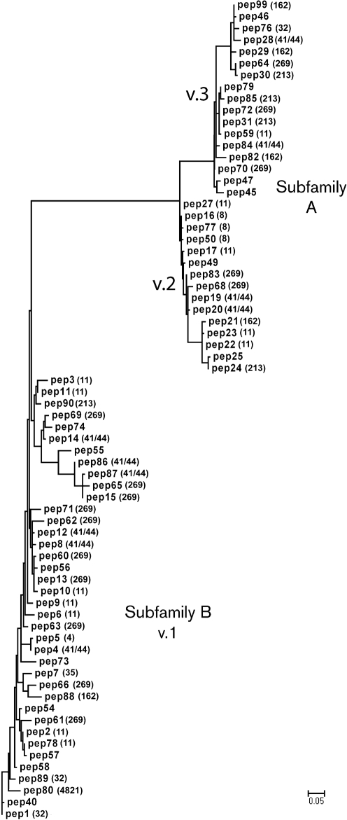
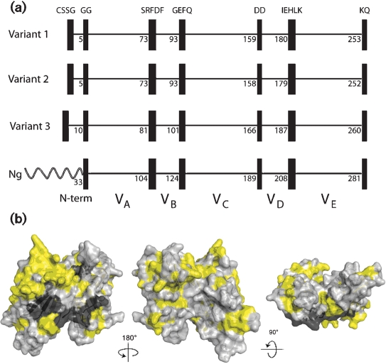
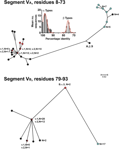
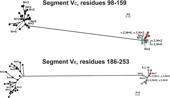
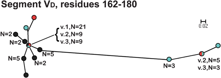
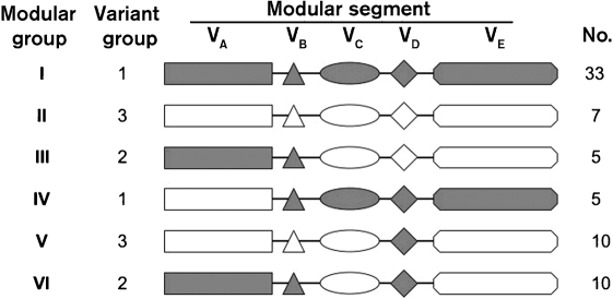
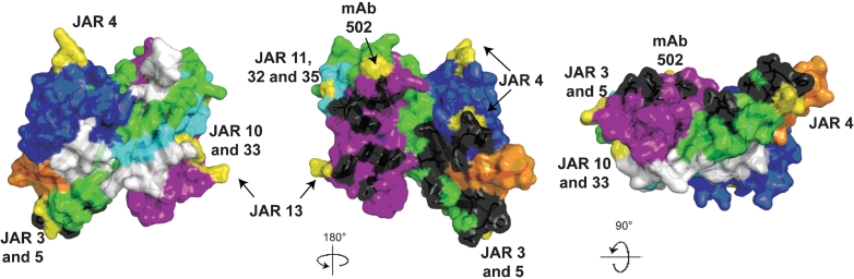
Meningococcal factor H binding protein (fHbp) is a promising vaccine antigen that binds the human complement downregulatory molecule factor H (fH), and this binding enhances the survival of the organism in serum. Based on sequence variability of the entire protein, fHbp has been divided into three variant groups or two subfamilies. Here, we present evidence based on phylogenetic analysis of 70 unique fHbp amino acid sequences that the molecular architecture is modular. From sequences of natural chimeras we identified blocks of two to five invariant residues that flanked five modular variable segments. Although overall, 46 % of the fHbp amino acids were invariant, based on a crystal structure, the invariant blocks that flanked the modular variable segments clustered on the membrane surface containing the amino-terminal lipid anchor, while the remaining invariant residues were located throughout the protein. Each of the five modular variable segments could be classified into one of two types, designated alpha or beta, based on homology with segments encoded by variant 1 or 3 fHbp genes, respectively. Forty of the fHbps (57 %) comprised only alpha (n=33) or beta (n=7) type segments. The remaining 30 proteins (43 %) were chimeras and could be classified into one of four modular groups. These included all 15 proteins assigned to the previously described variant 2 in subfamily A. The modular segments of one chimeric modular group had 96 % amino acid identity with those of fHbp orthologs in Neisseria gonorrhoeae. Collectively, the data suggest that recombination between Neisseria meningitidis and N. gonorrhoeae progenitors generated a family of modular, antigenically diverse meningococcal fHbps.


Figures

Fig. 1.
Fig. 1.
Phylogram of fHbp based on 70 unique amino acid sequences. For each sequence, the peptide identification number assigned in the fHbp peptide database at http://Neisseria.org is shown and, if known, the MLST clonal complex is shown in parentheses. The lower left branch shows variant group 1 as defined by Masignani et al. (2003) (subfamily B of Fletcher et al., 2004); subfamily A contained two branches, variant groups 2 and 3. The phylogram was constructed by multiple sequence alignment as described in Methods and Supplementary Table S2. Bar, five amino acid changes per 100 residues.
Fig. 2.
Fig. 2.
(a) Schematic representation of fHbp, showing positions of blocks of invariant residues (shown as black vertical rectangles). The top three panels show representative architectures of three N. meningitidis fHbp variants in groups 1, 2 and 3 (peptide ID nos 1, 16 and 28, respectively). The amino acid positions of the last residue in each variable segment are shown. With the exception of a longer, unrelated, amino-terminal element, two N. gonorrhoeae orthologues (Ng, GenBank accession nos AE004969 and CP001050) had the identical six invariant blocks of residues that flanked segments VA to VE. (b) Space-filling structural models of fHbp based on the coordinates of the protein in a complex with a fragment of human fH (Schneider et al., 2009). The light-grey residues represent the variable amino acids located within the modular variable segments. The invariant blocks of residues separating each of the variable segments are shown in dark grey. The yellow residues represent the invariant amino acids outside of these blocks. The model on the left is the surface predicted to be anchored to the cell wall. The model in the centre has been rotated 18 ° on the y axis from the corresponding model on the left, while the model on the right has been rotated 9 ° on the x axis as compared with the model in the centre. The figure was constructed with PyMol (http://www.pymol.org).
Fig. 3.
Fig. 3.
Phylograms of unique fHbp amino acid sequences in modular variable segments VA (residues 8–73) and VB (residues 79–93). Segments derived from fHbp in the variant group 1 are shown as black circles, the variant 2 group as red circles, and the variant 3 group as light-blue circles. Where multiple proteins possessed an identical sequence in a segment, the number of proteins is given. A.β.9 and B.α.3 refer to proteins with exceptional junctional points (see text). Bar, two amino acid changes per 100 residues. The histogram (Segment VA) shows the mean number of protein sequences (y axis) and percentage amino acid identity (x axis) generated by comparing each of the 48 α A segments with the corresponding α- and β-type A segments of all 70 proteins. The corresponding histograms comparing each of the α segments with the corresponding α and β types, and each of the β segments with the corresponding α and β types, for all five modular variable segments, VA to VE, are shown in Supplementary Fig. S1.
Fig. 4.
Fig. 4.
Phylograms of unique fHbp amino acid sequences in variable segments VC (residues 98–159) and VE (residues 186–253). The colours of the circles for segments in each variant group correspond to those described in the legend of Fig. 3. E.β.10 refers to peptide ID no. 82 with an exceptional junctional point (see text). Bars, two amino acid changes per 100 residues.
Fig. 5.
Fig. 5.
Phylogram of unique fHbp amino acid sequences in variable segment VD (residues 162–180). The colours of the circles for peptides in each variant group correspond to those described in the legend of Fig. 3. Bar, two amino acid changes per 100 residues.
Fig. 6.
Fig. 6.
Schematic representation of six fHbp modular groups deduced from phylogenetic analysis. Forty of the 70 proteins contained only α-type segments or β-type segments, and were designated modular groups I and II, respectively. The remaining 30 proteins were chimeras with different combinations of α (shown in grey) and β segments (shown in white), and could be assigned to one of four modular groups (III–VI). The relationship between the modular group and Masignani variant group designation, and the number of unique sequences observed within each fHbp modular group, are shown.
Fig. 7.
Fig. 7.
Space-filling structural models of fHbp based on the coordinates of the protein in a complex with a fragment of human fH (Schneider et al., 2009). The five variable segments VA to VE are depicted in different colours (VA, blue; VB, orange; VC, green; VD, light blue; VE, violet) and the invariant blocks of residues separating each of the variable segments are shown in white. The model in the middle was generated by rotation of 18 ° around the y axis relative to the respective model on the far left. The model on the right was generated by a 9 ° rotation around the x axis relative to the model in the middle. The fH contact residues are depicted in black, and the residues affecting the epitopes of anti-fHbp mAbs are shown in yellow. The figure was constructed with PyMol (http://www.pymol.org).

References

    1. Arreaza, L., Alcala, B., Salcedo, C., de la Fuente, L. & Vazquez, J. A. (2003). Dynamics of the penA gene in serogroup C meningococcal strains. J Infect Dis 187, 1010–1014. - PubMed
    1. Bambini, S., Muzzi, A., Olcen, P., Rappuoli, R., Pizza, M. & Comanducci, M. (2009). Distribution and genetic variability of three vaccine components in a panel of strains representative of the diversity of serogroup B meningococcus. Vaccine 27, 2794–2803. - PubMed
    1. Beernink, P. T. & Granoff, D. M. (2008). Bactericidal antibody responses induced by meningococcal recombinant chimeric factor H-binding protein vaccines. Infect Immun 76, 2568–2575. - PMC - PubMed
    1. Beernink, P. T., Welsch, J. A., Harrison, L. H., Leipus, A., Kaplan, S. L. & Granoff, D. M. (2007). Prevalence of factor H-binding protein variants and NadA among meningococcal group B isolates from the United States: implications for the development of a multicomponent group B vaccine. J Infect Dis 195, 1472–1479. - PMC - PubMed
    1. Beernink, P. T., Welsch, J. A., Bar-Lev, M., Koeberling, O., Comanducci, M. & Granoff, D. M. (2008). Fine antigenic specificity and cooperative bactericidal activity of monoclonal antibodies directed at the meningococcal vaccine candidate, factor H-binding protein. Infect Immun 76, 4232–4240. - PMC - PubMed

Publication types

MeSH terms

LinkOut - more resources