. 2009 Sep;111(1):37-48.
doi: 10.1093/toxsci/kfp139. Epub 2009 Jul 2.

Quantitative determination of skin penetration of PEG-coated CdSe quantum dots in dermabraded but not intact SKH-1 hairless mouse skin

Affiliations

Quantitative determination of skin penetration of PEG-coated CdSe quantum dots in dermabraded but not intact SKH-1 hairless mouse skin

Neera V Gopee et al. Toxicol Sci. 2009 Sep.

Abstract

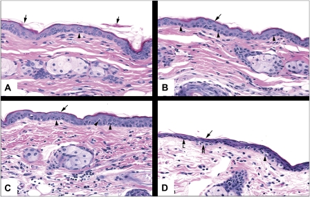
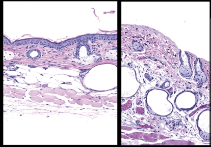
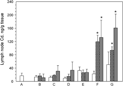
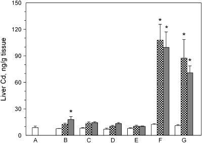
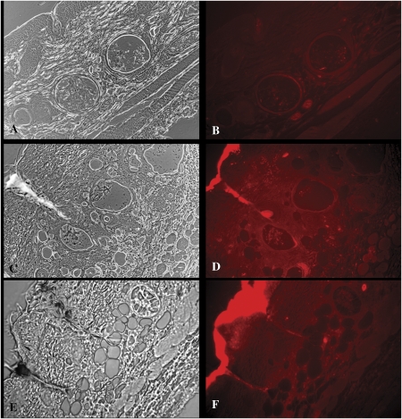
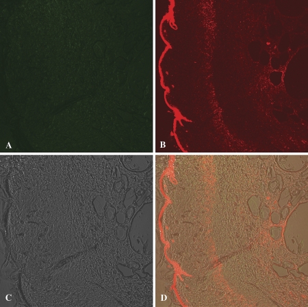
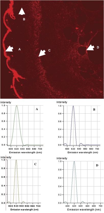
Many cosmetics, sunscreens, and other consumer products are reported to contain nanoscale materials. The possible transdermal absorption of nanoscale materials and the long-term consequences of the absorption have not been determined. We used polyethylene glycol coated cadmium selenide (CdSe) core quantum dots (QD; 37 nm diameter) to evaluate the penetration of nanoscale material into intact, tape stripped, acetone treated, or dermabraded mouse skin. QD were suspended in an oil-in-water emulsion (approximately 9 microM) and the emulsion was applied at 2 mg/cm(2) to mouse dorsal skin pretreated as follows: intact; tape stripped to remove the stratum corneum; acetone pretreated; dermabraded to remove stratum corneum and epidermis. QD penetration into the skin was monitored in sentinel organs (liver and regional draining lymph nodes) using inductively coupled plasma mass spectrometry analysis of cadmium (from the CdSe QD). No consistent cadmium elevation was detected in the sentinel organs of mice with intact, acetone pretreated, or tape-stripped skin at 24- and 48-h post-QD application; however, in dermabraded mice, cadmium elevations were detected in the lymph nodes and liver. QD accumulation (as cadmium) in the liver was approximately 2.0% of the applied dose. The passing of QD through the dermabraded skin was confirmed using confocal fluorescence microscopy. These results suggest that transdermal absorption of nanoscale materials depends on skin barrier quality, and that the lack of an epidermis provided access to QD penetration. Future dermal risk assessments of nanoscale materials should consider key barrier aspects of skin and its overall physiologic integrity.


Figures

FIG. 1.
FIG. 1.
Photomicrographs of skin from SKH-1 hairless mice following the application and removal of tape strips 0 (A), 5 (B), 10 (C), and 15 (D) times. The skin was stained with hematoxylin and eosin. The arrows point to partially desquamated keratin, and the arrow heads refer to cells in the stratum basale with perinuclear halos. Magnification is at ×20.
FIG. 2.
FIG. 2.
Dermabrasion was used to remove the stratum corneum and epidermis. The panel on the left is skin from an untreated mouse. The skin section on the right is from a mouse where the epidermis was removed using dermabrasion (1 h prior). The skin was stained with hematoxylin and eosin. Magnification is at ×20.
FIG. 3.
FIG. 3.
Cd levels in regional lymph nodes of SKH-1 mice topically applied with CdSe QD suspended at 9μM in an oil-in-water emulsion. The axial and brachial lymph nodes were removed from mice (n = 3 per group) topically treated as follows: (A) no QD applied; (B) QD applied to normal skin; (C) QD applied to tape-stripped skin; (D) QD applied to acetone treated skin; (E) QD applied to untreated skin and covered with occlusion patch; (F) QD applied to dermabraded skin; (G) QD applied to dermabraded skin and covered with occlusion patch. The animals were sacrificed at 0 h (open bar), 24 h (hatched bar), or 48 h (filled bar) after application of the emulsion.
FIG. 4.
FIG. 4.
Cd levels in the liver of SKH-1 mice topically applied with CdSe QD suspended at 9μM in an oil-in-water emulsion. The liver was removed from mice (n = 3 per group) topically treated as follows: (A) no QD applied; (B) QD applied to normal skin; (C) QD applied to tape-stripped skin; (D) QD applied to acetone treated skin; (E) QD applied to untreated skin and covered with occlusion patch; (F) QD applied to dermabraded skin; (G) QD applied to dermabraded skin and covered with occlusion patch. The animals were sacrificed at 0 h (open bar), 24 h (hatched bar), or 48 h (filled bar) after application of the emulsion.
FIG. 5.
FIG. 5.
Photomicroscopic bright field (A, C, E) and fluorescence (B, D, F) images of intact skin (A, B), and dermabraded skin with (C, D) and without an occlusion patch (E, F) 48 h after topical application of QD. The epidermis is in the upper left corner of each image. The QD, which emit fluorescence at 621 nm, appear red in the images.
FIG. 6.
FIG. 6.
Representative images generated by confocal fluorescence microscopy are shown of dermabraded skin treated for 24 h with QD. The upper left image (A) was taken using a band-pass filter (505–550 nm) allowing collection of autofluorescence, whereas the upper right image (B) was taken using a long-pass filter (> 575 nm) which would contain the target QD (fluorescence emission at 621 nm). The lower left panel (C) shows the differential interference contrast image (Nomarski interference contrast) image of the skin, and the lower right image (D) is a composite of images (A–C).
FIG. 7.
FIG. 7.
The fluorescence spectra in the indicated areas (A–D) of the representative tissue sample were determined using the spectrum analysis component of the confocal fluorescence microscope. The spectra for each of the points (A–D) indicated in the figure are shown in the lower spectral plots. Point-A is of QD on the stratum corneum, and had maximum fluorescence at 620 nm. Similarly, fluorescence in areas (B–D) in the dermis had spectra indicative of the 621-nm QD.

References

    1. Alekseenko AV, Waseem TV, Fedorovich SV. Ferritin, a protein containing iron nanoparticles, induces reactive oxygen species formation and inhibits glutamate uptake in rat brain synaptosomes. Brain Res. 2008;1241:193–200. - PubMed
    1. Arakaki A, Nakazawa H, Nemoto M, Mori T, Matsunaga T. Formation of magnetitie by bacteria and its application. J. R. Soc. Interface. 2008;5:977–999. - PMC - PubMed
    1. Bailey R. 2003. The smaller the better: the limitless promise of nanotechnology and the prowing peril of a moratorium. ReasonOnline. Available at: www.reason.com/news/show/28969.html. Accessed March 13, 2009.
    1. Baroli B, Ennas MG, Loffredo F, Isola M, Pinna R, López-Quintela MA. Penetration of metallic nanoparticles in human full-thickness skin. J. Invest. Dermatol. 2007;127:1701–1712. - PubMed
    1. Bhattacharya P, Ghosh S, Stiff-Roberts AD. Quantum dot opto-electronic devices. Annu. Rev. Mater. Res. 2004;34:1–40.

Publication types