. 2009 Aug;175(2):547-56.
doi: 10.2353/ajpath.2009.081011. Epub 2009 Jul 2.

Macrophages are alternatively activated in patients with endometriosis and required for growth and vascularization of lesions in a mouse model of disease

Affiliations

Macrophages are alternatively activated in patients with endometriosis and required for growth and vascularization of lesions in a mouse model of disease

Monica Bacci et al. Am J Pathol. 2009 Aug.

Abstract

The mechanisms that sustain endometrial tissues at ectopic sites in patients with endometriosis are poorly understood. Various leukocytes, including macrophages, infiltrate endometriotic lesions. In this study, we depleted mouse macrophages by means of either clodronate liposomes or monoclonal antibodies before the injection of syngeneic endometrial tissue. In the absence of macrophages, tissue fragments adhered and implanted into the peritoneal wall, but endometriotic lesions failed to organize and develop. When we depleted macrophages after the establishment of endometriotic lesions, blood vessels failed to reach the inner layers of the lesions, which stopped growing. Macrophages from patients with endometriosis and experimental mice, but not nonendometriotic patients who underwent surgery for uterine leiomyomas or control mice, expressed markers of alternative activation. These markers included high levels of scavenger receptors, CD163 and CD206, which are involved in both the scavenging of hemoglobin with iron transfer into macrophages and the silent clearance of inflammatory molecules. Macrophages in both inflammatory liquid and ectopic lesions were equally polarized, suggesting a critical role of environmental cues in the peritoneal cavity. Adoptively transferred, alternatively activated macrophages dramatically enhanced endometriotic lesion growth in mice. Inflammatory macrophages effectively protected mice from endometriosis. Therefore, endogenous macrophages involved in tissue remodeling appear as players in the natural history of endometriosis, required for effective vascularization and ectopic lesion growth.


Figures
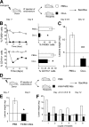
Figure 1
Figure 1
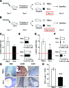
Peritoneal macrophages are required for endometriosis establishment. A: Estradiol benzoate-treated female mice were sacrificed and uteri removed and split. Endometrial tissue was isolated and mechanically disrupted before intraperitoneal injection of an experimental/control mice pair. Control mice (open squares) and depleted mice (filled squares) were treated i.p. at day 0, 4, and 8 with liposomes containing PBS (PBS-L) or clodronate (Clo-L) respectively. Lesions were evaluated 12 days after endometrial tissue injection. B: The percentage of peritoneal F4/80+ and of CD11b+ macrophages was evaluated by flow cytometry at different time points (left panels). Right panels represent the quantitative expression of the results obtained at day 12, expressed as percentage of positive cells in mice treated with PBS-L (open columns) or Clo-L (filled columns) respectively. C: The dry weight of endometriotic lesions from mice treated with PBS-L or with Clo-L was assessed at day 12. Results are expressed as mean of total weights of lesions from six independent experimental/control mouse pair ± SEM. D: Estradiol benzoate-treated female mice were sacrificed and uteri removed and split. Endometrial tissue was isolated and mechanically disrupted before intraperitoneal injection of experimental/control mice pairs. Mice were treated with PBS (open squares) or F4/80 mAb mice (filled squares) i.p. 2 days before injection of syngeneic endometrial tissue (day −2) and every other day since then (days 0, 2, 4, 6, 8, 10, and 12). Lesions were evaluated at day 12, when mice were sacrificed. E: The dry weight of endometriotic lesions from mice treated with PBS (open columns) or with F4/80 mAb (filled columns) was assessed at day 12. Results are expressed as mean of total weights of lesions from six independent experimental/control mouse pair ± SEM. F: The dry weight of endometriotic lesions from mice treated with PBS or with F4/80 mAb was assessed at day 12. Results from each independent experimental/control mouse pair, in which endometrial tissue from a single donor was split and injected in a mouse previously injected with PBS (open columns) or with F4/80 mAb (filled columns). Significantly different from control, *P < 0.05 and **P < 0.01.
Figure 2
Figure 2
Peritoneal macrophages sustain the growth of established endometriotic lesions. Estradiol benzoate-treated female mice were sacrificed and uteri removed and split. Endometrial tissue was isolated and mechanically disrupted before i.p. injection of an experimental/control mice pair. Control mice (open squares) and depleted mice (filled squares) were treated i.p. with liposomes containing PBS (PBS-L) or clodronate (Clo-L) 24 hours before (A) or 4 and 8 days after (B) i.p. injection of syngeneic endometrial tissue. Lesions were evaluated at day 12 day after endometrium injection. The dry weight of endometriotic lesions from mice treated with PBS-L (open columns) or with Clo-L (filled columns) 24 hours before (C) or 4 and 8 days after (E) i.p. injection of syngeneic endometrial tissue was assessed at day 12. Results are expressed as mean of total weights of lesions from six independent experimental/control mouse pair±SEM. F4/80+ and CD11b+ macrophages were identified by flow cytometry in animals treated with PBS-L or Clo-L 24 hours before (D) or 4 and 8 days after (F) i.p. injection of syngeneic endometrial tissue at sacrifice at day 12. Panels report the results expressed as percentage of peritoneal positive cells in mice treated with PBS-L (open columns) or Clo-L (filled columns). G: Macrophages (CD68+ cells) and endothelial cells (CD31+ cells) in the endometriotic lesions obtained from mice treated with PBS-L or with Clo-L were evaluated by immunohistochemistry at day 12. H: The treatment effect on vascularization was also quantitatively assessed by counting the number of CD31+ cells per field of vision. Results represent the mean ± SEM of five independent field of vision, as assessed by two independent blinded expert pathologists. Significantly different from control, *P < 0.05 and ***P < 0.001.
Figure 3
Figure 3
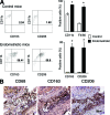
Peritoneal macrophages of mice with established endometriotic lesion express markers of alternative activation. A: Peritoneal cells were retrieved at day 12 from mice injected either with PBS (control mice, open symbols) of from mice 12 days after injection of syngeneic endometrial tissue (endometriotic mice, filled symbols), as described in Materials and Methods. Left plots: CD11b+ cells (y axis, red fluorescence) were analyzed by flow cytometry for the expression of markers of alternative activation, such as the CD163 and the CD206 antigens (x axis, green fluorescence). Right panels: histograms represent the fraction of peritoneal cells expressing each marker in control (open columns) or endometriotic (filled columns) mice. Results represent the mean ± SEM of three independent experiments. B: Endometrial lesions were characterized by immunohistochemistry for the expression of CD68, CD163, and CD206 in infiltrating macrophages.
Figure 4
Figure 4
Peritoneal macrophages of endometriotic patients express markers of alternative activation. A: Peritoneal cells from endometriotic patients (filled columns) and relevant controls (open columns) were analyzed for the expression of macrophage lineage, activation, and differentiation markers by flow cytometry. The cluster analyzed was defined based on the expression of the CD45 common leukocyte antigen (y axis, red fluorescence) by cells with appropriate side scatter characteristics (x axis, green fluorescence) as illustrated. B: % of peritoneal macrophages expressing the indicated markers in endometriotic patients (filled columns) and relevant controls (open columns). Results are expressed as the mean ± SEM of 23 endometriotic patients and 13 matched controls. C: Expression of indicated markers by peritoneal macrophages from endometriotic patients (filled columns) and relevant controls (open columns) as assessed by the associated relative fluorescence intensity (y axis) calculated as described in Materials and Methods). Results are expressed as the mean ± SEM of 23 endometriotic patients and 13 matched controls. Significantly different from control, *P < 0.05 and **P < 0.01.
Figure 5
Figure 5
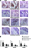
Macrophages infiltrating endometriotic lesions and apparently healthy peritoneum express markers of alternative activation. A: Macrophages in endometriotic lesions, leiomyomas, and apparently nonaffected peritoneum from patients with endometriosis and controls were characterized by immunohistochemistry for the expression of a lineage marker (CD68) and for markers of alternative activation (CD163 and CD206). B: Infiltrating macrophages expressing CD68, CD163, and CD206 were quantitatively assessed by the infiltration score, performed as described in Materials and Methods. *P < 0.05, significantly different from control.
Figure 6
Figure 6
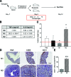
Endometriotic lesions growth is dependent on macrophages activation. A: Estradiol benzoate-treated female mice were sacrificed and uteri were removed and split. Endometrial tissue was isolated and mechanically disrupted before i.p. injection of four experimental/control mice pair per each treatment group. Control mice were treated with PBS. Experimental animals were injected i.p. at day −2, 0, and + 2 with M0, M1, and M2 macrophages (1.5 × 10⁶cells/mouse/100 μl). Animals were sacrificed 12 days after endometrial tissue injection. B: TNF-α and IL-10 secretion by in vitro polarized M0, M1, and M2 macrophages was assessed by enzyme-linked immunosorbent assay. Results are expressed as the mean ± SEM of at least three independent experiments. C: The dry weight of endometriotic lesions was assessed in mice treated with PBS or injected with M0, M1, or M2 macrophages. Results are expressed as mean of total weights of lesions from at leas four mice ± SEM. *P < 0.05, significantly different from control group. D: Representative H&E staining and immunohistochemical analysis for CD68+ cells in endometriotic lesions obtained from mice treated with M1 and M2 macrophages.

References

    1. Lebovic DI, Mueller MD, Taylor RN. Immunobiology of endometriosis. Fertil Steril. 2001;75:1–10. - PubMed
    1. Giudice LC, Kao LC. Endometriosis. Lancet. 2004;364:1789–1799. - PubMed
    1. Berkley KJ, Rapkin AJ, Papka RE. The pains of endometriosis. Science. 2005;308:1587–1589. - PubMed
    1. Sampson J. Peritoneal endometriosis due to menstrual dissemination of endometrial tissue into the peritoneal cavity. Am J Obstet Gynecol. 1927;14:422–469.
    1. Dunselman GA, Groothuis PG. Etiology of endometriosis: hypotheses and facts. Gynecol Obstet Invest. 2004;57:42–43. - PubMed

Publication types