. 2009 Aug;175(2):835-43.
doi: 10.2353/ajpath.2009.090095. Epub 2009 Jul 2.

Development of arterial blood supply in experimental liver metastases

Affiliations

Development of arterial blood supply in experimental liver metastases

Katalin Dezso et al. Am J Pathol. 2009 Aug.

Abstract

In this study, we present a mechanism for the development of arterial blood supply in experimental liver metastases. To analyze the arterialization process of experimental liver metastases, we elucidated a few key questions regarding the blood supply of hepatic lobules in mice. The microvasculature of the mouse liver is characterized by numerous arterioportal anastomoses and arterial terminations at the base of the lobules. These terminations supply one hepatic microcirculatory subunit per lobule, which we call an arterial hepatic microcirculatory subunit (aHMS). The process of arterialization can be divided into the following steps: 1) distortion of the aHMS by metastasis; 2) initial fusion of the sinusoids of the aHMS at the tumor parenchyma interface; 3) fusion of the sinusoids located at the base of the aHMSs, which leads to the disruption of the vascular sphincter (burst pipe); 4) incorporation of the dilated artery and the fused sinusoids into the tumor; and 5) further development of the tumor vasculature (arterial tree) by proliferation, remodeling, and continuous incorporation of fused sinusoids at the tumor-parenchyma interface. This process leads to the inevitable arterialization of liver metastases above the 2000- to 2500-mum size, regardless of the origin and growth pattern of the tumor.


Figures

Figure 1
Figure 1
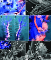
A: Liver lobes after the casting procedure containing arterial (red) and mixed blood (white) metastases of the C38 tumor line. The blue resin fills only the terminal portal venules. B: The same lobes after corrosion. Arrows point to the hollow spaces of metastases not having arterial blood supply. Arrowhead points to a metastasis of which only the base was filled by the red resin; therefore its arterial connection was undetectable on the uncorroded specimen (arrowhead on A). Scale bar = 3 mm.
Figure 2
Figure 2
Normal mouse liver. A: Direct arterioportal anastomosis (arrow) on a large trunk of the portal tree. A small amount of the red resin is spread on the previously hardened blue resin. Scale bar = 1 mm. B: Detail of A (arrowhead in A) viewed under the scanning electron microscope. Anastomosis between the peribiliary plexus and the portal vessel. Numerous capillaries of the peribiliary plexus (arrowhead) joined into one vessel that enters the large trunk of the portal vein (arrow). Scale bar = 300 μm. C and D: Detail of a vascular tree of the liver viewed from opposite directions. The portal vein is filled with blue resin. Scale bar for C,D = 500 μm. C: Shows the side where the artery (arrowhead) is running. Note the numerous aHMSs (arrows) spaced regularly between terminal portal venules (marked by arrows on D) along the portal vein. Each space between the terminal portal venules corresponds to one lobulus. D: No aHMSs are visible on the other side of the portal vein. E: Light micrograph of a single aHMS located above the peribiliary plexus and between terminal portal venules (arrows) at the base of the lobule. The space between the terminal portal venules determines the extension of the lobule. The red resin, which fills the sinusoids of the aHMS is in connection (arrowheads) with blue resin of the terminal portal venules, showing that the aHMS is part of the lobule. Scale bar = 200 μm. F,G: An arterial HMS at the periphery of the vascular tree of the liver, SEM images. F: The arteriole (large arrowhead) runs up on the portal venule and terminates in an aHMS at the base of the lobule (small arrow). The lobule is defined by the tree terminal portal venules (small arrowheads). Note that the red resin (inset) is present in the central venule (large arrow). The inset shows light micrograph of the same area. Scale bar = 200 μm. G: High power micrograph of the aHMS shown on Figure 2F. The main branch of the arteriole terminates in the aHMS (arrow). Smaller branches form direct anastomoses (arrowheads) with the portal venule. Scale bar = 90 μm.
Figure 3
Figure 3
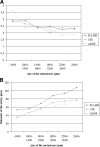
Percentage of arterial metastases in relation to metastasis size.
Figure 4
Figure 4
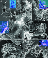
A: A C38 micrometastasis on the surface of the liver grows close to a terminal portal venule (small arrowheads). The vascular lake (arrows) at the surface of the metastasis is filled with blue resin (inset) injected through the portal vein. An arteriole (large arrowhead) is visible close to the ramification of the portal venule, which later represents the base of the lobule. The metastasis has not yet reached this region. Scale bar = 500 μm; 500 μm (inset). B: Detail of A. The arteriole branched off (arrows) is close the fork of the terminal portal venule. The resin hardly entered the aHMS (arrowheads). Scale bar = 90 μm. C: Impression of a C 38 metastasis in an aHMS (small arrowheads). The sinusoids of the aHMS are nearly normal in structure. Inset shows the same aHMSs filled with red resin (arrow) at the base of the metastasis and the supplying arteriole (large arrowhead). Broken line marks the border of the metastasis. Small arrowheads point at the terminal portal venules entrapped within the metastasis. Scale bars: 100 μm; 200 μm (inset). D: Compressed aHMS at the base (inset) of a C38 metastasis. Initial fusion of the sinusoids of the HMS is discernible (arrows). Other sinusoids of the aHMS are normal in structure (small arrows). On the inset the arrow points to an arteriole supplying the HMS. Drops of the resin (red dot on the light micrograph marked by arrowhead) are present in the central venule (arrowheads). Scale bars: 90 μm; 300 μm (inset). E: The “nest” of a metastasis viewed from the top (from the surface of the liver). The sinusoids of the aHMS at the base of the metastasis are partially fused (arrowheads). Arrow points at a portal vessel within the metastasis projecting toward the surface of the liver. Scale bar = 100 μm. F: Fusion of sinusoids in the metastasis of the 3LL-HH tumor. Large vascular lakes are not formed at the surface; instead tortuous vessels appear within the metastasis (large arrowheads). Note the impressions left by small tissue pillars (arrows) within the tumor vessels representing the last step of the fusion (reverse intussusceptive angiogenesis). The low density tortuous tumor vessels are continuous with the high density sinusoids of the surrounding liver tissue. Scale bar = 60 μm. G: Small A2058 metastasis. The centrally located non-dilated artery (arrow) ramifies into delicate intratumoral vessels. Scale bars: 200 μm; 500 μm (inset).
Figure 5
Figure 5
A,B: Formation of a vascular lake from an aHMS at surface of the metastasis of the C38 tumor line. A: The arrow points to the non-dilated arteriole feeding the severely distorted and fused aHMS (arrowheads). A broken line is drawn along two terminal portal venules that are pushed aside by the growing tumor mass and thereby outlines the border of the metastasis. Scale bar = 500 μm. B: The area marked by arrowheads on A is viewed from above by the scanning electron microscope. Extensive fusion of the sinusoids leads to vascular lake formation (arrowheads). The arrow points to the supplying arteriole. Scale bar = 200 μm. C: Part of an arterialized C38 micrometastasis. The supplying arteriole is severely dilated (arrow) and ramifying into vessels, which delineate the surface (arrowheads) of the metastasis. The arteriole has not yet been incorporated into the metastasis. Scale bar = 100 μm. D: Scanning electron micrograph of the same structure shown on (C) viewed from above. Fused sinusoids (arrows) and vessels (arrowheads) are organized into basket-like form. Scale bar = 200 μm. E: Arterialized C38 micrometastasis. Dilated arteriole (arrow) feeds the metastasis. The area where most of the branches arise (arrowheads) is just incorporated into the tumor but the vessels run on or close to the surface of the metastasis. The center of the metastasis is avascular. Scale bar = 200 μm. F: 3LL-HH metastasis supplied by a strongly dilated artery (arrow). There is an extreme size difference between the artery running toward the tumor and the artery accompanying the neighboring portal vein (arrowhead). Note that the two portal veins are about the same size. Scale bar = 1 mm.
Figure 6
Figure 6
A: C38 metastasis with a well developed central arterial tree (black arrowheads on the inset). The portal vein was pushed aside (arrows). No ramifications can be observed on the SEM micorgraph along the trunk of the arterial tree (white arrowheads). Scale bars: 600 μm; 1 mm (inset). B: Detail of A. Vascular branches originate from one small area of the arterial tree (arrow) located in the center of the metastasis. Scale bar = 300 μm. C: 3LL-HH metastasis. The centrally located artery dilates gradually toward the center of the metastasis where it ends in tortuous vascular lakes. The metastasis is not completely filled by the resin its borders are defined by the portal branches (arrowheads). Scale bars: 600 μm; 500 μm (inset). D: C38 metastasis with a strongly dilated funnel-like artery (arrow). Both the artery and the portal vein (arrowhead) are located centrally. The portal vein is extremely compressed obstructing the resin flow. Note that there are no ramifications along the trunk of the artery. The branching point of the artery is located in the center of the metastasis. Scale bars: 200 μm; 500 μm (inset).
Figure 7
Figure 7
A: Portal vein-artery ratio in relation to metastasis size. B: Diameter of the supplying artery at the entry to the metastasis in relation to metastasis size.
Figure 8
Figure 8
Schematic representation of the arterialization process in liver metastases. Step 0: Micrometastasis growing within the liver lobule. The arterial HMS has normal architecture. Step 1: Distortion of the aHMS by the metastasis. Step 2: Initial fusion of the sinusoids of the aHMSs at the tumor parenchyma interface. Step 3: Fusion of the sinusoids located at the base of the HMSs, leading to the disruption of the sphincter (burst pipe). Step 4: Incorporation of the dilated artery and the fused sinusoids. Step 5: Further development of the tumor vasculature (arterial tree) by proliferation, remodeling, and continuous incorporation of fused sinusoids at the surface of the tumor. Blue - mixed portal and arterial blood, Purple - mixed blood in the arterial HMSs, Red - arterial blood, Green - hepatocytes, Gray - tumor tissue, Black boxes - arterial sphincters.

References

    1. Breedis C, Young G. The blood supply of neoplasms in the liver. Am J Pathol. 1954;30:969–977. - PMC - PubMed
    1. Archer SG, Gray BN. Vascularization of small liver metastases. Br J Surg. 1989;76:545–548. - PubMed
    1. Lin G, Lunderquist A, Hägerstrand I, Boijsen E. Postmortem examination of the blood supply and vascular pattern of small liver metastases in man. Surgery. 1984;96:517–526. - PubMed
    1. Ackerman NB. The blood supply of experimental liver metastases. IV Changes in vascularity with increasing tumor growth. Surgery. 1974;75:589–596. - PubMed
    1. Ridge JA, Bading JR, Gelbard AS, Benua RS, Daly JM. Perfusion of colorectal hepatic metastases. Relative distribution of flow from the hepatic artery and portal vein. Cancer. 1987;59:1547–1553. - PubMed

Publication types