. 2009 Oct 15;180(8):701-12.
doi: 10.1164/rccm.200901-0100OC. Epub 2009 Jul 2.

Deletion of Pten expands lung epithelial progenitor pools and confers resistance to airway injury

Affiliations

Deletion of Pten expands lung epithelial progenitor pools and confers resistance to airway injury

Caterina Tiozzo et al. Am J Respir Crit Care Med. .

Abstract

Rationale: Pten is a tumor-suppressor gene involved in stem cell homeostasis and tumorigenesis. In mouse, Pten expression is ubiquitous and begins as early as 7 days of gestation. Pten(-/-) mouse embryos die early during gestation indicating a critical role for Pten in embryonic development.

Objectives: To test the role of Pten in lung development and injury.

Methods: We conditionally deleted Pten throughout the lung epithelium by crossing Pten(flox/flox) with Nkx2.1-cre driver mice. The resulting Pten(Nkx2.1-cre) mutants were analyzed for lung defects and response to injury.

Measurements and main results: Pten(Nkx2.1-cre) embryonic lungs showed airway epithelial hyperplasia with no branching abnormalities. In adult mice, Pten(Nkx2.1-cre) lungs exhibit increased progenitor cell pools composed of basal cells in the trachea, CGRP/CC10 double-positive neuroendocrine cells in the bronchi, and CC10/SPC double-positive cells at the bronchioalveolar duct junctions. Pten deletion affected differentiation of various lung epithelial cell lineages, with a decreased number of terminally differentiated cells. Over time, Pten(Nxk2.1-cre) epithelial cells residing in the bronchioalveolar duct junctions underwent proliferation and formed uniform masses, supporting the concept that the cells residing in this distal niche may also be the source of procarcinogenic stem cells. Finally, increased progenitor cells in all the lung compartments conferred an overall selective advantage to naphthalene injury compared with wild-type control mice.

Conclusions: Pten has a pivotal role in lung stem cell homeostasis, cell differentiation, and consequently resistance to lung injury.


Figures
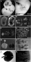
Figure 1.
Figure 1.
Nkx2.1-cre expression during lung development. (AK) Detection of cre-induced β-galactosidase activity at different embryonic stages. (A, B) E10.5 whole mount β-galactosidase staining of Rosa26RNkx2.1-cre detecting (A) activity at the level of the brain and the lung primordia (arrows). (B) Notice the strongest staining at the airway level (arrow). (C, D) β-galactosidase staining of WT and RosaR26Nkx2.1-cre embryos at E13.5. (C) The control does not present any staining, whereas (D) the Rosa26RNkx2.1-cre shows staining at the level of the brain (arrow), (E) thyroid (arrow), and (D) lung (arrow). (EG) At E13.5, Lac-Z expression in the distal lung is heterogeneous, with areas more stained compared with others. The extrapulmonary and intrapulmonary airways were labeled completely, whereas the distal parenchyma presents some spots with a decreased degree of activity (G, higher magnification of F). (H, I) At E15.5, the majority of the cells were labeled in both of the compartments. (I) Vibrotome section through E15.5 lung. (J, K) At the adult stage, the majority of the cells in the distal compartment are stained. The airways present always a strongest β-galactosidase activity compared with the distal compartment (K, higher magnification of J). Br = brain; L = lung; T = thyroid.
Figure 2.
Figure 2.
Deletion of Pten does not affect branching morphogenesis during lung development. (AD) Hematoxylin and eosin (H&E) staining of lung sections of control animals (n = 4, A and C) and mutant PtenNkx2.1-cre (n = 4, B and D) at E15.5, detecting no differences in branching between the two groups. Magnification A and B, ×10; C and D, ×20. (EH) Lung sections were stained with PTEN antibodies and NKX2.1 antibodies (magnification ×40); in the control, the cells (G) expressing NKX2.1 also (E) expressed PTEN. In the mutant, (H) these cells did not present PTEN staining, except (F) for very few cells (arrows). (I, J) H&E staining of trachea sections of wild type (n = 4, I) and mutant (n = 4, J) at E15.5, showing the epithelium hyperplasia present in the PtenNkx2.1-cre trachea. (K, L) Trachea sections were stained with PTEN antibodies; in the control the cells were positive for PTEN, whereas (L) in the mutant there was an homogenous deletion of the gene in all the epithelial cells. (M) Tissue-specific deletion of Pten was also proved by polymerase chain reaction analysis. Primers for recombination analysis were designed as described previously (22). P1/P2 amplified the floxed and the wt allele, when the P1/P3 amplified the flanked-exon 5 (Δ5). e = epithelium, br = bronchi.
Figure 3.
Figure 3.
Absence of Pten leads to bronchiolar hyperplasia secondary to an increase in proliferation rate and to a decrease in apoptosis. (AD) Histological analysis through hematoxylin and eosin staining of lungs from wild type and mutants at E15.5 and E18.5 embryonic stage showing the presence of the epithelial hyperplasia. (E, F) E-cadherin staining for epithelial cells in the adult stage (PN60). (GJ) Ki67 staining in PN60 lungs detecting an increase of the Ki67-positive cells number in the PtenNkx2.1cre mice compared with the control (G and H, magnification ×20; I and J, magnification ×80). (K, L) TUNEL (terminal deoxynucleotidyl transferase dUTP nick end labeling) assay in the mutant and control lungs. (M) Quantification revealed a statistically significant difference between the two groups (n = 3 mice per genotype), *P ≤ 0.01 using the standard t test. (N) Statistical analysis of the apoptotic cells (n = 3 mice for genotype). *P ≤ 0.01 using the standard t test. Lm = lumen.
Figure 4.
Figure 4.
Deletion of Pten increases number of basal cells in the PtenNkx2.1-cre trachea. (AJ) Lung sections from mutant PtenNkx2.1-cre and control littermates at 2 months of age were stained (A and B) for P63 and (C and D) for keratin14. Increased number of double-positive cells over the P63-positive cells was detected in the mutant lungs compared with control lungs (low magnification, G and H; high magnification, I and J). (K) Quantification analysis was performed using t test from three mice in each group, *P ≤ 0.01.
Figure 5.
Figure 5.
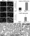
Inactivation of Pten leads to an increase of the neuroepithelial bodies in the bronchi. (AH) Immunofluorescence for CC10 (A and B) and CGRP (C and D) in 8-week-old mutant and control lung sections. Increased size and brightness in the neuroepithelial bodies (NEB) (G and H, arrows) were observed in the PtenNkx2.1-cre lungs compared with the control animals. (I) Quantification analysis. The average and standard deviation from four mice were compared using the t test, *P ≤ 0.01. (J) Relative expression of CGRP mRNA in the PtenNkx2.1-cre and control mice, confirming the statistically significant increase of the CGRP expression in the mutant compared with the control (data from three different animals, P ≤ 0.05). (K, L) Immunohistochemistry for CGRP in E18.5 control and mutant lungs, showing an increase of NEB numbers already during embryonic stages. Similar results are obtained in adult stages. Lm = lumen.
Figure 6.
Figure 6.
Deletion of Pten increases the double-positive cells CC10/SPC in the BADJ. (A, D) Hematoxylin and eosin staining showed in the mutant an increase of cells at the BADJ level (compare A to B, lower magnification ×20; C to D, higher magnification, ×40) and these cells were also enlarged compared with the cells in the control (D, arrows). (EJ) Immunofluorescence for SPC and CC10 in control (n = 3) and mutant PtenNkx2.1cre (n = 3) animals at PN60: the mutants presented an increased number of double-positive cells (E, F, and G, lower magnification, ×20; H, I, and J, higher magnification, ×80). (KN) Double immunofluorescence was also detected in the mutant using a confocal microscope to confirm the staining in single double-positive cells (arrows). (O) Quantification analyses were performed in three mice from each group using the t test, *P < 0.01. (P, Q) Fluorescence-activated cell sorter analysis of control (n = 3) and mutant (n = 3) lungs detecting CD45CD31CD34+Sca-1+ cells.
Figure 7.
Figure 7.
The BADJ cells proliferate inside the parenchyma and give rise to masses. (A, B) Hematoxylin and eosin staining on PN180 PtenNkx2.1-cre lung showing that over time the cells in the BADJ grow in the parenchyma and give rise to a tumorlike mass. (C) E-cadherin staining showed that the tumor cells are epithelial cells acting as progenitors, forming structures (underscored) that resemble the branching happening during the pseudoglandular stage. (D, E) Immunofluorescence for SPC and CC10 detected double-positive cells (arrows) in the mass. (F) N-myc staining showed an increase of N-myc nuclear staining in tumor cells. Underscored line delimits the masse from the surrounding parenchyma. (G, H) Immunohistochemistry for Ki67 inside the masses showing very few cells positive for Ki67 (G). Statistical analysis did not show any statistically significant difference between the masses and the surrounding parenchyma (H). m = mass; p = parenchyma.
Figure 8.
Figure 8.
Deletion of Pten increases p-AKT and β-catenin activation. (A, B) Immunofluorescence for β-cateninSer522 and E-cadherin in E15.5 lung sections. Double-positive cells are increased in the mutant (B) compared with the control (A). (C, D) Immunohistochemistry for phospho-AKT in E15.5 lung sections: the PtenNkx2.1-cre lungs (D) showed up-regulation of phospho-AKT compared with the control (C). Pictures are representative of n = 3 separate mice per genotype, P ≤ 0.01. (E) Statistical analysis of double-positive cells for β-cateninSer522 and E-cadherin double-positive cells, showing the statistically significant increase of double-positive cells in the mutant compared with the control (n = 3 animals for each group). (G) Immunoblot for phospho-AKT in extract of whole lungs taken from control and mutants at 3 weeks of age, confirming the immunohistochemistry data. Data are representative of two animals for each group.
Figure 9.
Figure 9.
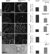
Absence of Pten impairs cell fate. (AH) Immunofluorescence for (A, B) CC10, (C, D) β-tubulin, (E, F) SPC, and (G, H) T-1α from 2-month-old control and mutant lungs. (A, B) In the mutant, an increase of Clara cells (CC10 positive) is detected compared with the control (B). (C, D) β-tubulin staining showed a reduction in the ciliated cell number in the mutant (C, arrows) compared with the control (D). (E, F) Alveolar type II cells (SPC positive) were increased in the PtenNkx2.1-cre (E). (G, H) Decrease in alveolar type I cells (T-1 α positive) was observed in the mutant. (I, J) Immunohistochemistry for HES-1 in 2-month-old control and mutant lung. (KN) Related expression, as determined by real-time polymerase chain reaction of (I) CC10, (J) Tubulin IV, (K) SPC, and (L) Aqp-5 mRNAs in PtenNkx2.1-cre and control mice confirmed the lack of differentiation in the mutant lung (data from three different mice in each group). (O, P) Related expression of Hes-1 mRNA in PtenNkx2.1-cre and control mice confirmed the increase of HES-1 expression in the mutant. *P ≤ 0.05.
Figure 10.
Figure 10.
Absence of Pten protects the airways from naphthalene injury. (AF) Hematoxylin and eosin staining of a control and mutant trachea after corn oil or naphthalene injection. (A, D) Corn oil administration did not affect the tracheal structure in the control (A) or in the mutant (D). (B, E) Three days after naphthalene administration, tracheal epithelium in the control was completely denuded (B), whereas the PtenNkx2.1-cre tracheal epithelium did not show any sign of injury and the cells were able to survive (E). (C, F) At 7 days after injury, the Ptenf/f trachea showed a reepithelization (C), whereas the mutant did not show any change in the morphology (F). (GL) Immunofluorescence for CC10 in the bronchi. (G, J) Control (G) and mutant (J) bronchi did not present any damage after corn oil administration. (H, K) At 3 days after naphthalene injury, the injury in the control (H) was more extensive and severe compare with the mutant (K). (I, L) After 7days, the PtenNkx2.1-cre (L) presented more cells compared to the control (I). Data from four different animals for each group. (M) Morphometric quantification of the epithelial damage at Day 1, 3, and 7 after naphthalene injury. Epithelial damage was measured as a ratio between nude surface and total surface in bronchi. Ten different fields from control and mutant lungs were considered (n = 4 animals in each group). *P ≤ 0.05. (N) Quantification of the double-positive cells for SPC/CC10 versus total number of CC10-positive cells in control and mutant lungs at 1 day after injury. *P ≤ 0.05. (O) Statistical analysis of the double-positive cells for PH3/CC10 versus total number of CC10-positive cells in the control and mutant lungs at 7 days after injury (n = 4). *P ≤ 0.05. lm = lumen; p = parenchyma.

Similar articles

Cited by

References

    1. Fuchs E, Tumbar T, Guasch G. Socializing with the neighbors: stem cells and their niche. Cell 2004;116:769–778. - PubMed
    1. Lanza R. Essentials of stem cells biology. New York: Academic Press; 2006.
    1. Engelhardt JF, Schlossberg H, Yankaskas JR, Dudus L. Progenitor cells of the adult human airway involved in submucosal gland development. Development 1995;121:2031–2046. - PubMed
    1. Schoch KG, Lori A, Burns KA, Eldred T, Olsen JC, Randell SH. A subset of mouse tracheal epithelial basal cells generates large colonies in vitro. Am J Physiol Lung Cell Mol Physiol 2004;286:L631–L642. - PubMed
    1. Avril-Delplanque A, Casal I, Castillon N, Hinnrasky J, Puchelle E, Péault B. Aquaporin-3 expression in human fetal airway epithelial progenitor cells. Stem Cells 2005;23:992–1001. - PubMed

Publication types