

Endosomal TLR signaling is required for anti-nucleic acid and rheumatoid factor autoantibodies in lupus
- PMID: 19574451
- PMCID: PMC2715524
- DOI: 10.1073/pnas.0905441106
Endosomal TLR signaling is required for anti-nucleic acid and rheumatoid factor autoantibodies in lupus
Abstract
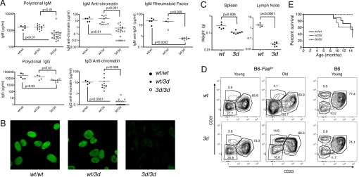
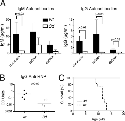
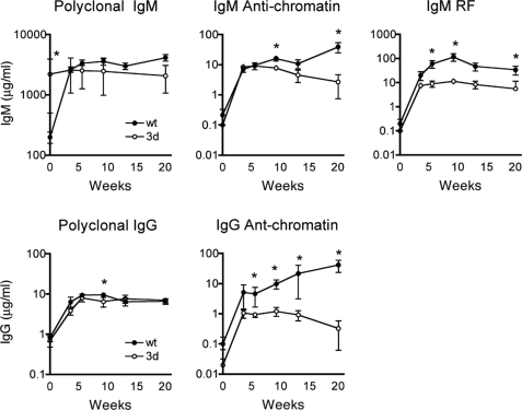
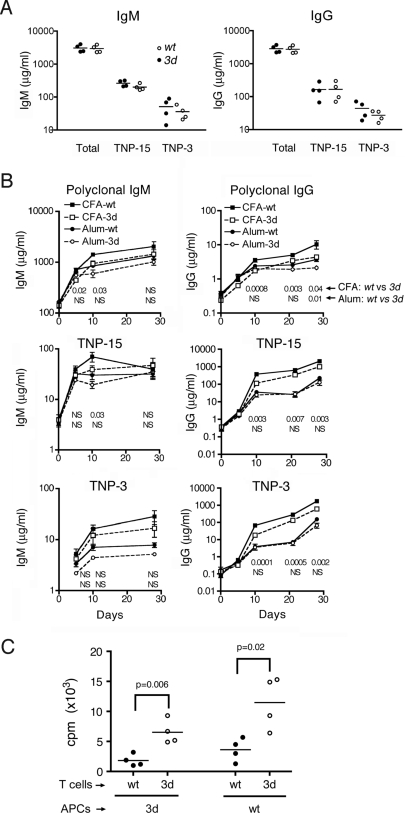
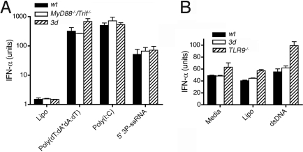
Using the Unc93b1 3d mutation that selectively abolishes nucleic acid-binding Toll-like receptor (TLR) (TLR3, -7, -9) signaling, we show these endosomal TLRs are required for optimal production of IgG autoAbs, IgM rheumatoid factor, and other clinical parameters of disease in 2 lupus strains, B6-Fas(lpr) and BXSB. Strikingly, treatment with lipid A, an autoAb-inducing TLR4 agonist, could not overcome this requirement. The 3d mutation slightly reduced complete Freund's adjuvant (CFA)-mediated antigen presentation, but did not affect T-independent type 1 or alum-mediated T-dependent humoral responses or TLR-independent IFN production induced by cytoplasmic nucleic acids. These findings suggest that nucleic acid-sensing TLRs might act as an Achilles' heel in susceptible individuals by providing a critical pathway by which relative tolerance for nucleic acid-containing antigens is breached and systemic autoimmunity ensues. Importantly, this helps provide an explanation for the high frequency of anti-nucleic acid Abs in lupus-like systemic autoimmunity.
Conflict of interest statement
The authors declare no conflict of interest.
Figures
References
-
- Arbuckle MR, et al. Development of autoantibodies before the clinical onset of systemic lupus erythematosus. N Engl J Med. 2003;349:1526–1533. - PubMed
-
- Reveille JD. Predictive value of autoantibodies for activity of systemic lupus erythematosus. Lupus. 2004;13:290–297. - PubMed
-
- Vlahakos DV, et al. Anti-DNA antibodies form immune deposits at distinct glomerular and vascular sites. Kidney Int. 1992;41:1690–1700. - PubMed
-
- Marshak-Rothstein A, Rifkin IR. Immunologically active autoantigens: The role of toll-like receptors in the development of chronic inflammatory disease. Annu Rev Immunol. 2007;25:419–441. - PubMed
Publication types
MeSH terms
Substances
Grants and funding
- GM67759/GM/NIGMS NIH HHS/United States
- R01 GM067759/GM/NIGMS NIH HHS/United States
- R01 AR039555/AR/NIAMS NIH HHS/United States
- AI059777/AI/NIAID NIH HHS/United States
- AR39555/AR/NIAMS NIH HHS/United States
- R37 GM067759/GM/NIGMS NIH HHS/United States
- AR053228/AR/NIAMS NIH HHS/United States
- R01 AR031203/AR/NIAMS NIH HHS/United States
- R37 AR039555/AR/NIAMS NIH HHS/United States
- R01 ES007511/ES/NIEHS NIH HHS/United States
- AR31203/AR/NIAMS NIH HHS/United States
- R01 AR053228/AR/NIAMS NIH HHS/United States
- R01 AR053731/AR/NIAMS NIH HHS/United States
- ES07511/ES/NIEHS NIH HHS/United States
- AR42242/AR/NIAMS NIH HHS/United States
- R01 AR042242/AR/NIAMS NIH HHS/United States
- R01 AI059777/AI/NIAID NIH HHS/United States
LinkOut - more resources
Full Text Sources
Other Literature Sources
Medical
Molecular Biology Databases
Research Materials
Miscellaneous
