

Maturation of thalamic radiations between 34 and 41 weeks' gestation: a combined voxel-based study and probabilistic tractography with diffusion tensor imaging
- PMID: 19574497
- PMCID: PMC7051519
- DOI: 10.3174/ajnr.A1660
Maturation of thalamic radiations between 34 and 41 weeks' gestation: a combined voxel-based study and probabilistic tractography with diffusion tensor imaging
Abstract
Background and purpose: This study aimed to investigate brain maturation along gestational age with diffusion tensor imaging in healthy preterm and term neonates. Therefore, a voxel-based study of fractional anisotropy (FA) and mean diffusivity (D(av)) was performed to reveal the brain regions experiencing microstructural changes with age. With tractography, the authors intended to identify which fiber tracts were included in these significant voxels.
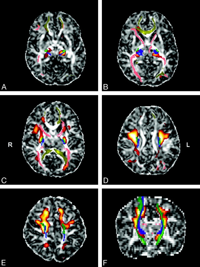
Materials and methods: There were 22 healthy preterm and 6 healthy term infants who underwent MR imaging between 34 and 41 weeks of gestation. A statistical parametric approach was used to evidence the effect of age on regional distribution of FA and D(av) values. The fiber tracts suspected to be included in the significant clusters of voxels were identified with neuroanatomy and tractography atlases, reconstructed with probabilistic tractography, and superimposed on the parametric maps.
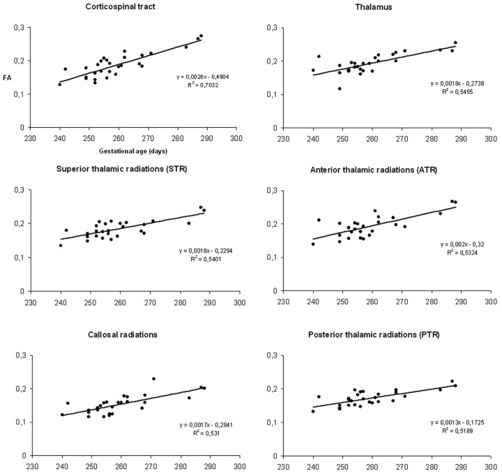
Results: Parametric analysis showed that FA increases with age in the subcortical projections from the frontal (motor and premotor areas) and parietal cortices, the centrum semiovale, the anterior and posterior arms of the internal capsules, the optic radiations, the corpus callosum, and the thalami (P < .05, corrected). Superimposition of the parametric maps on tractography showed that the corticospinal tract (CST); the callosal radiations (CR); and the superior, anterior, and posterior thalamic radiations were included in the significant voxels. No statistically significant results were found for D(av) maps.
Conclusions: These results highlight that, besides the already-evidenced FA increase in the CST and CR, the thalami and the thalamic radiations experience microstructural changes in the early development of the human brain.
Figures

References
-
- Sidman RL, Rakic P. Development of the human nervous system. In: Haymaker W, Adams R.D, ed. Histology and Histopathology of the Nervous System. Springfield: Charles C. Thomas, 1982:3–145
-
- Paus T, Collins DL, Evans AC, et al. Maturation of white matter in the human brain: a review of magnetic resonance studies. Brain Res Bull 2001;54:255–66 - PubMed
-
- Huppi PS, Dubois J. Diffusion tensor imaging of brain development. Semin Fetal Neonatal Med 2006;11:489–97 - PubMed
-
- Basser PJ, Pierpaoli C. Microstructural and physiological features of tissues elucidated by quantitative-diffusion-tensor MRI. J Magn Reson B 1996;111:209–19 - PubMed
Publication types
MeSH terms
LinkOut - more resources
Full Text Sources