. 2009 Aug 19;28(16):2469-78.
doi: 10.1038/emboj.2009.184. Epub 2009 Jul 2.

Insights into complement convertase formation based on the structure of the factor B-cobra venom factor complex

Affiliations

Insights into complement convertase formation based on the structure of the factor B-cobra venom factor complex

Bert J C Janssen et al. EMBO J. .

Abstract

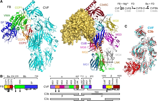
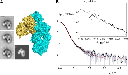
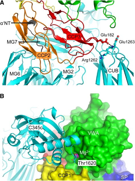
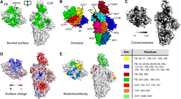
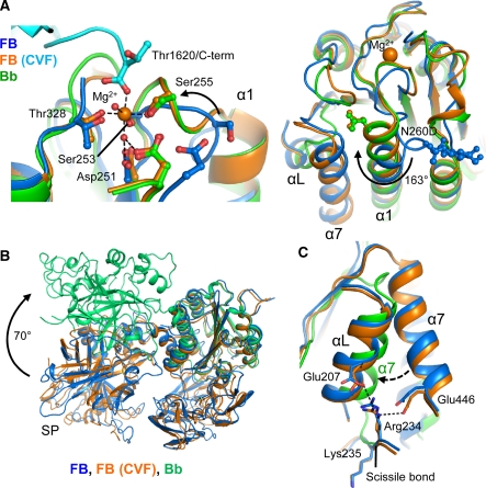
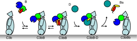
Immune protection by the complement system critically depends on assembly of C3 convertases on the surface of pathogens and altered host cells. These short-lived protease complexes are formed through pro-convertases, which for the alternative pathway consist of the complement component C3b and the pro-enzyme factor B (FB). Here, we present the crystal structure at 2.2-A resolution, small-angle X-ray scattering and electron microscopy (EM) data of the pro-convertase formed by human FB and cobra venom factor (CVF), a potent homologue of C3b that generates more stable convertases. FB is loaded onto CVF through its pro-peptide Ba segment by specific contacts, which explain the specificity for the homologous C3b over the native C3 and inactive products iC3b and C3c. The protease segment Bb binds the carboxy terminus of CVF through the metal-ion dependent adhesion site of the Von Willebrand factor A-type domain. A possible dynamic equilibrium between a 'loading' and 'activation' state of the pro-convertase may explain the observed difference between the crystal structure of CVFB and the EM structure of C3bB. These insights into formation of convertases provide a basis for further development of complement therapeutics.


Conflict of interest statement

The authors declare that they have no conflict of interest.

Figures

Figure 1
Figure 1
Structure of the CVFB complex at 2.2-Å resolution. (A) Ribbon representation of CVFB with FB coloured by domain and CVF coloured cyan (left) and of CVF coloured by domain with FB in wheat surface representation (right). The proteolytic assembly process of the C3 convertase is shown schematically. (B) Domain compositions, including disulphide bridges and glycan positions, of FB and CVF are indicated, together with the topology of C3b and C3c for clarity. (C) Comparison of CVF (cyan) with C3b (Janssen et al, 2006) (red) (see also Supplementary Table IIA).
Figure 2
Figure 2
The CVFB crystal structures correlate well with EM and SAXS of CVFB in solution. (A) EM class averages of the pro-convertase CVFB (I–III) correlate well with the crystal structure of CVFB, shown in surface representation (cyan and wheat, respectively, with the FB scissile bond black) and as a low-resolution projection (P). (B) The computed scattering curve of the 2.2-Å crystal structure of CVFB (red line) fitted with CRYSOL (Svergun et al, 1995) to the measured scattering data of 2 mg/ml CVFB (black dots with experimental errors) with good correlation (χ2 of 1.2). The computed scattering curves of the 3.0-Å crystal structures of CVFB gave good correlation (both χ2 of 1.2) with the measured scattering data as well (not shown). Inset; Guinier plot for CVFB from X-ray scattering.
Figure 3
Figure 3
The CVFB interface consists of two patches. (A) Ribbon representation of CCP2–3 (coloured orange and red, respectively) of the Ba segment interacting with MG2, MG6, MG7, CUB (all coloured cyan) and α′NT (coloured black) of CVF. Glu182 of FB interacts with Arg1262 and Glu1263 of CVF (ball-and-stick representation), which correspond to the first FI-cleavage site in C3b. (B) VWA of the Bb segment, shown in green surface representation, interacts with C345C of CVF shown in cyan ribbon representation. The C-terminus (Thr1620) of CVF binds to the Mg2+ ion (purple sphere) in FB.
Figure 4
Figure 4
Surface representation of the CVFB interface coloured functionally. (A) An opened view of the 4900 Å2 footprint of the FB–CVF interface is highlighted in green. (B) Domains of FB and CVF coloured according to Figure 1. (C) FB and CVF colour-coded to residue conservation; from non-conserved (white) to conserved (black). Figure is produced using CONSURF (Glaser et al, 2005). (D) FB and CVF coloured by electrostatic potential from red (−10 kbT/ec) to blue (−10 kbT/ec). The VWA:C345C interface consists of conserved complementary electrostatic patches. (E) Previously proposed sites involved in complex formation. The yellow coloured patches are epitopes to which antibody binding decreases complex formation (Hourcade et al, 1995; Thurman et al, 2005). The other patches are based on FB to C2 chimeras that increase binding of FB to C3b >150% (Hourcade et al, 1995) (blue) or decrease binding <10% (Tuckwell et al, 1997) (green); on C3 to CVF chimeras that increase C3bBb complex stability (Kolln et al, 2005; Fritzinger et al, 2009) (lime); on an alternative proteolytic product of C3, that supports activation of FB (O'Keefe et al, 1988) (orange) or on single site mutants (single numbers) that increase complex formation (Hourcade et al, 1999) (dark red) or decrease complex formation (Taniguchi-Sidle and Isenman, 1994) (red). Legend for colour-coding and residue numbers are presented in the table. CVF residue numbering is according to human C3.
Figure 5
Figure 5
Conformational rearrangements in the VWA domain of FB. (A) The MIDAS site in VWA rearranges from distorted in free FB (Milder et al, 2007) (blue) to a high-affinity Mg2+-bound conformation in CVFB (cyan and orange) similar to free Bb (Ponnuraj et al, 2004) (green) (left panel). Helix α1 elongates and glycan-linked Asn260 (mutated to Asp in CVFB) rotates 163° (right panel). (B) Comparison of free FB (blue), FB bound to CVF (orange) and free Bb (lime). (C) Helixes αL and α7 and the Arg234–Lys235 scissile bond do not rearrange on FB binding to CVF. Arg234 remains hydrogen bonded to Glu207 and Glu446. In Bb α7 has swapped with αL that is removed. Colour scheme as in (A).
Figure 6
Figure 6
Convertase formation and activation model. Schematic representation of the assembly of the pro-convertase complex and its subsequent activation by FD into the active C3 convertase (colour scheme as in Figure 1A, left panel).

References

    1. Adams PD, Grosse-Kunstleve RW, Hung LW, Ioerger TR, McCoy AJ, Moriarty NW, Read RJ, Sacchettini JC, Sauter NK, Terwilliger TC (2002) PHENIX: building new software for automated crystallographic structure determination. Acta Crystallogr D Biol Crystallogr 58: 1948–1954 - PubMed
    1. Bhattacharya AA, Lupher ML Jr, Staunton DE, Liddington RC (2004) Crystal structure of the A domain from complement factor B reveals an integrin-like open conformation. Structure (Camb) 12: 371–378 - PubMed
    1. CCP4 (1994) The CCP4 suite: programs for protein crystallography. Acta Crystallogr D Biol Crystallogr 50: 760–763 - PubMed
    1. Durocher Y, Perret S, Kamen A (2002) High-level and high-throughput recombinant protein production by transient transfection of suspension-growing human 293-EBNA1 cells. Nucleic Acids Res 30: E9. - PMC - PubMed
    1. Emsley P, Cowtan K (2004) Coot: model-building tools for molecular graphics. Acta Crystallogr D Biol Crystallogr 60: 2126–2132 - PubMed

Publication types

MeSH terms