. 2009 Oct;16(10):1408-17.
doi: 10.1038/cdd.2009.86. Epub 2009 Jul 3.

Cyclin D1 promotes anchorage-independent cell survival by inhibiting FOXO-mediated anoikis

Affiliations

Cyclin D1 promotes anchorage-independent cell survival by inhibiting FOXO-mediated anoikis

L Gan et al. Cell Death Differ. 2009 Oct.

Erratum in

  • Cell Death Differ. 2010 May;17(5):900

Abstract

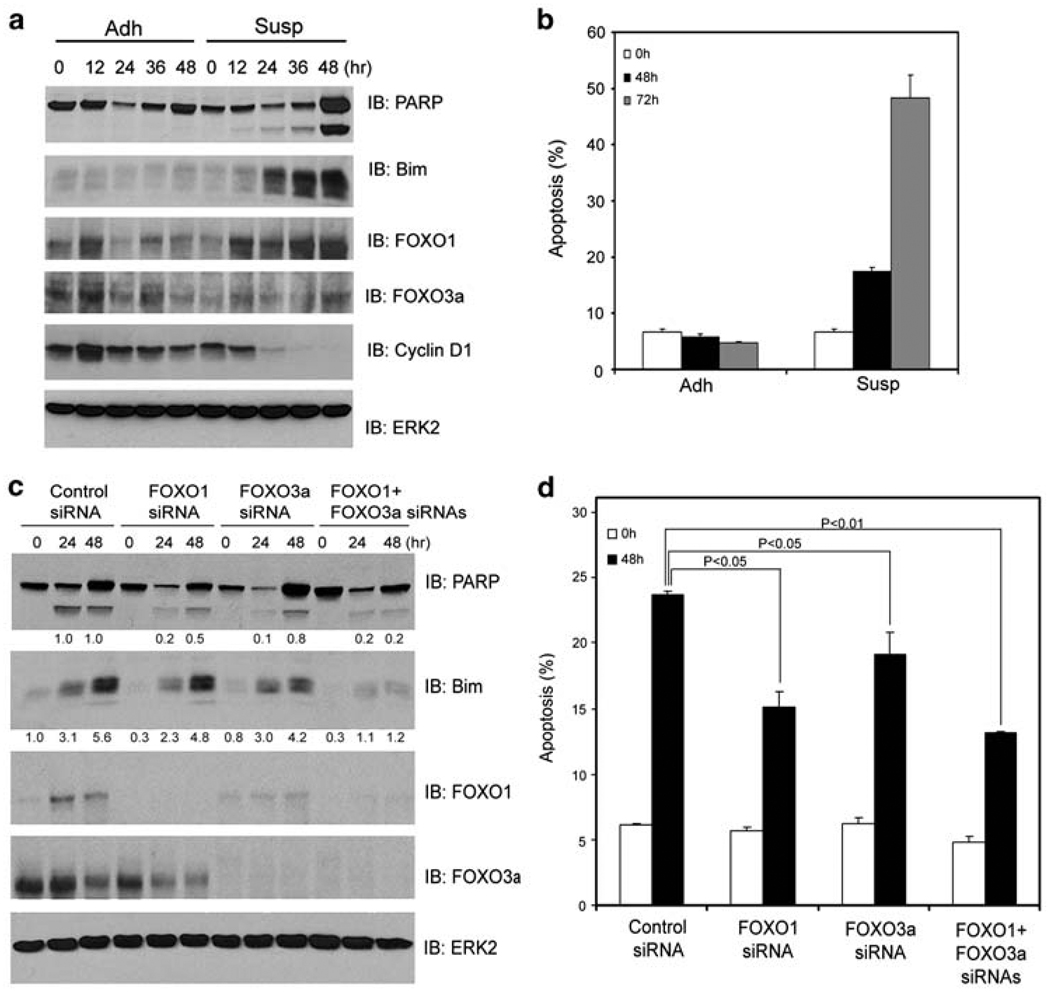
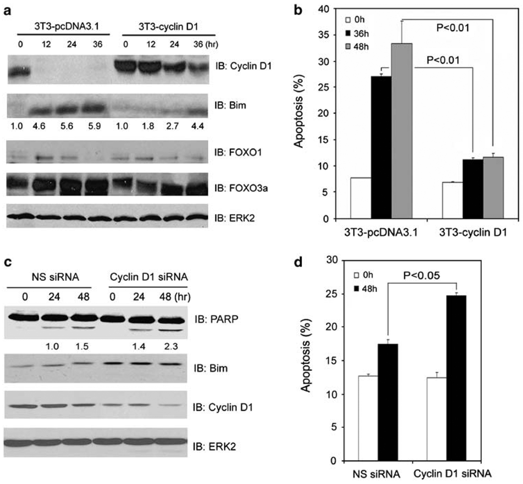
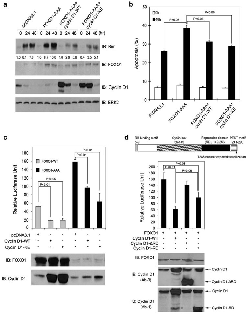
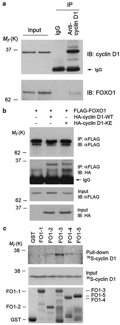
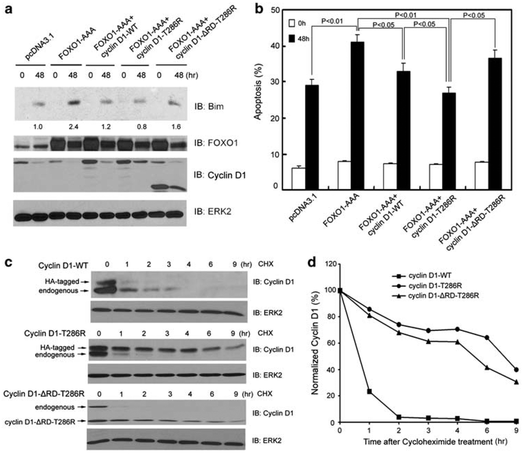
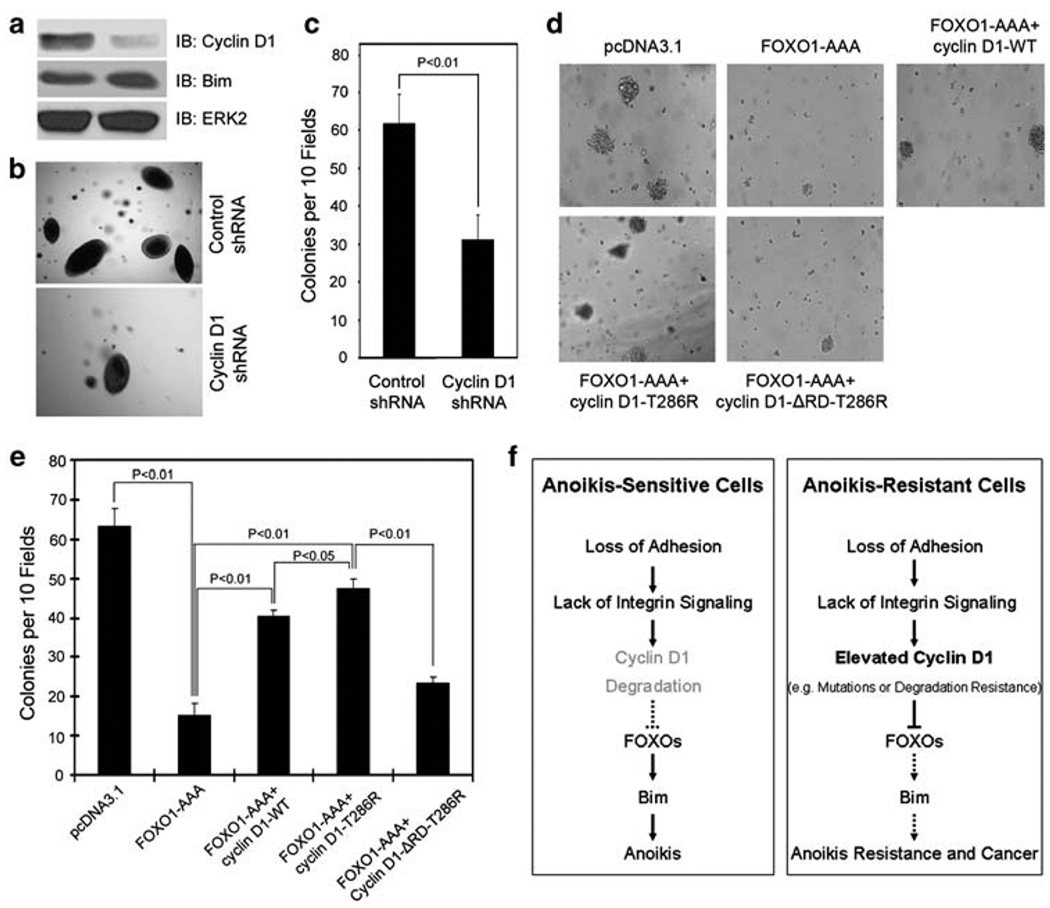
O-class forkhead box (FOXO) transcription factors are critical regulators of diverse cellular processes, including apoptosis, cell-cycle arrest, DNA damage repair and oxidative stress resistance. Here, we show that FOXO1 and FOXO3a have an essential function in promoting cell detachment-induced anoikis, resistance to which is implicated in cancer development and metastasis. In contrast, the oncoprotein cyclin D1 inhibits anoikis. We further show that cyclin D1 interacts with FOXO proteins and impedes their transcriptional regulatory and anoikis-promoting functions. This effect of cyclin D1 requires its transcription repression domain but is independent of cyclin-dependent kinases CDK4 and CDK6. Moreover, we show that cancer-derived mutants of cyclin D1 are much more stable than wild-type cyclin D1 under anchorage-independent conditions and possess a greater antagonistic effect on FOXO-regulated anoikis and anchorage-independent growth of cancer cells. These data suggest that cyclin D1 may have a critical function in tumorigenesis and cancer metastasis by inhibiting the anoikis-promoting function of FOXO proteins.


Conflict of interest statement

All authors claim no conflict of interest with this study.

Figures

Figure 1
Figure 1
FOXO1 and FOXO3a promote anoikis. DU145 cells grown in adhesion (Adh) or suspension (Susp) were collected at the indicated time points and subjected to immunoblotting analysis for expression of PARP, Bim, FOXO1, FOXO3a, and cyclin D1 proteins (a) and measurement of apoptosis (b). (c, d) DU145 cells were transfected with control siRNA, or siRNAs for FOXO1, FOXO3a, or both. At 48 h after transfection, cells were plated in adhesion or suspension and at the indicated time points cells were subjected to immunoblotting (c) and apoptosis (d) analysis. ERK2 was used as a loading control. Results from three independent experiments were quantified for apoptosis. Error bars indicate S.D. among three individual experiments
Figure 2
Figure 2
Cyclin D1 inhibits anoikis. (a, b) NIH3T3 cells stably expressing either an empty vector (3T3-pcDNA3.1) or cyclin D1 (3T3-cyclin D1) were cultured in suspension and at the indicated time points cells were subjected to immunoblotting (a) and apoptosis (b) analysis. (c, d) LNCaP cells were transfected with a pool of cyclin D1-specific siRNAs or nonspecific control siRNAs. At 48 h after transfection, cells were cultured in suspension and at the indicated time points cells were subjected to immunoblotting (c) and apoptosis (d) analysis. The number underneath each band in the immunoblot indicates the relative intensity of the corresponding band. Error bars indicate S.D. among three individual experiments
Figure 3
Figure 3
Cyclin D1 inhibits FOXO1-augmented anoikis and FOXO1’s transcriptional activity. (a, b) DU145 cells were transfected with plasmids as indicated. At 36 h after transfection, cells were cultured in suspension and at the indicated time points cells were subjected to immunoblotting (a) and apoptosis (b) analysis. The number underneath each band in the immunoblot indicates the relative intensity of the corresponding band. (c) LNCaP cells were transfected with firefly and Renilla luciferase reporter constructs and plasmids as indicated. At 36 h after transfection, cells were subjected to luciferase activity measurement as described in ‘Materials and Methods’ (upper panel) or western blot analysis (lower panel). Error bars indicate S.D. among three individual experiments. (d) Top, a schematic diagram of the cyclin D1 protein shows its different functional domains. Middle and bottom, LNCaP cells were transfected with the indicated plasmids and luciferase activities and western blots were analyzed as described in (c). The asterisk indicates a nonspecific immunoreactive band
Figure 4
Figure 4
Cyclin D1 interacts with FOXO1 in vitro and in vivo. (a) Interaction of endogenous cyclin D1 and FOXO1 proteins in LNCaP cells as shown by co-immunoprecipitation using an antibody against cyclin D1. (b) LNCaP cells were transfected with plasmids as indicated. At 48 h after transfection, cells were subjected to immunoprecipitation and western blot analysis. (c) Binding of 35S-labeled cyclin D1 proteins, produced by in vitro transcription and translation, to GST–FOXO1 recombinant proteins. Top, autography of in vitro-produced cyclin D1 proteins bound by GST–FOXO1 recombinant proteins. Middle, input of 35S-labeled cyclin D1 proteins. Bottom, GST and GST–FOXO1 recombinant proteins used for in vitro protein-binding assays indicated by Coomassie blue staining
Figure 5
Figure 5
Inhibitory effect on FOXO1-mediated anoikis and protein stability of wild type and a cancer-derived mutant of cyclin D1. (a, b) DU145 cells were transfected with plasmids as indicated. At 24 h after transfection, cells were cultured in suspension and at the indicated time points cells were subjected to immunoblotting (a) and apoptosis (b) analysis. The number underneath each band in the immunoblot indicates the relative intensity of the corresponding band. (c) DU145 cells were transfected with HA-tagged wild-type cyclin D1, HA-tagged cyclin D1-T286R, or cyclin D1-ΔRD-T286R mutants. At 48 h after transfection, DU145 cells were cultured in suspension and treated with 20 µg/ ml cycloheximide. Cells were collected at the indicated time points and cell lysates were analyzed by western blots. (d) Quantification of the cyclin D1 protein signal intensity was obtained from exposures in which the signal was not saturated during the entire time course. Signal intensities were normalized to the signal intensity obtained at time zero
Figure 6
Figure 6
Effect of the functional interaction between cyclin D1 and FOXO1 on anchorage-independent growth of cancer cells. (ac) 22Rv1 cells were transduced with lentivirus containing expression vectors for cyclin D1 shRNA or nonspecific control shRNA. At 3 days after infection, the cells were plated for growth in medium containing 0.3% agar. The remaining cells were used for western blot analysis (a). After 3 weeks, microscopic colonies were visualized and counted in a blinded fashion. Results from one representative field in each group are shown in (b). Quantification of results of colony formation data is presented in (c). Error bars indicate S.D. among three individual experiments. (d, e) LNCaP cells were transfected with plasmids as indicated. Transfected cells were plated for growth in medium containing 400 µg/ml of G418 and 0.3% agar. After 3 weeks, microscopic colonies were visualized and counted. Results from one representative field in each group are shown in (d). Quantification of results of colony formation data is presented in (e). Error bars indicate S.D. among three individual experiments. (f) A model of the role of cyclin D1 deregulation in tumorigenesis through sustained inhibition of FOXO1-mediated anoikis

References

    1. Schwartz MA. Integrins, oncogenes, and anchorage independence. J Biol Chem. 1997;139:575–578. - PMC - PubMed
    1. Frisch SM, Ruoslahti E. Integrins and anoikis. Curr Opin Cell Biol. 1997;9:701–706. - PubMed
    1. Giancotti FG, Ruoslahti E. Integrin signaling. Science. 1999;285:1028–1032. - PubMed
    1. Reginato MJ, Mills KR, Paulus JK, Lynch DK, Sgroi DC, Debnath J, et al. Integrins and EGFR coordinately regulate the pro-apoptotic protein Bim to prevent anoikis. Nat Cell Biol. 2003;5:733–740. - PubMed
    1. Mailleux AA, Overholtzer M, Schmelzle T, Bouillet P, Strasser A, Brugge JS. BIM regulates apoptosis during mammary ductal morphogenesis, and its absence reveals alternative cell death mechanisms. Dev Cell. 2007;12:221–234. - PMC - PubMed

Publication types