

A method for measuring the length of the cochlea through magnetic resonance imaging
- PMID: 19575114
- PMCID: PMC9450629
- DOI: 10.1016/s1808-8694(15)30788-6
A method for measuring the length of the cochlea through magnetic resonance imaging
Abstract
We know that hearing impairment affects a large part of the population. In cases of profound and bilateral hearing loss, children may have problems in speech development, as well as communication and socialization. Cochlear implants have been used as a treatment option in these cases. Today, inner ear MRI is a mandatory test in the preoperative evaluation of these individuals. In our daily routines, we wonder whether MRI can provide not only qualitative, but also quantitative data, with real cochlear linear values built from three dimension images.
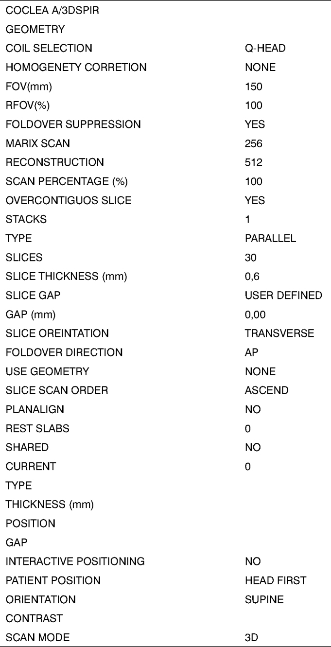
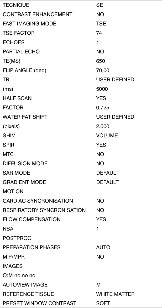
Aims: The aim of the present investigation is to propose a method to obtain MRI cochlear length measures from the temporal bones of cadavers.
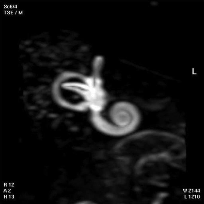
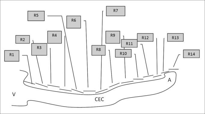
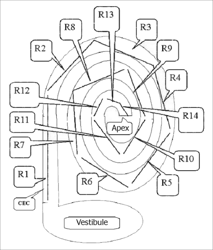
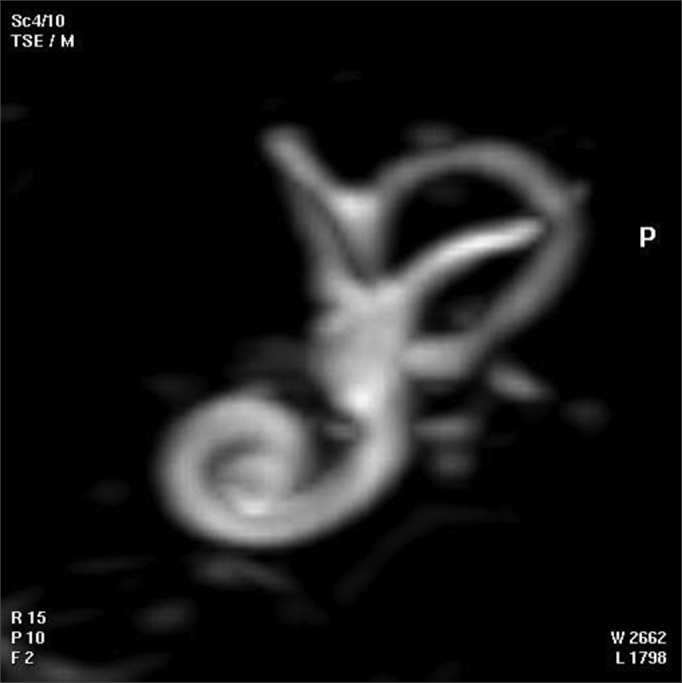
Material and methods: We assessed three dimensional images from the cochlea of six cadavers. By overlapping digitalized rulers on these images it was possible to measure cochlear length.
Results: These measures varied between 17 and 26.5 millimeters.
Conclusions: We have concluded that it was possible to measure cochlear length from three dimensional MRI images, by employing the method hereby proposed.
Figures
Similar articles
-
High-resolution computed tomography-based length assessments of the cochlea--an accuracy evaluation.Acta Otolaryngol. 2014 Oct;134(10):1011-5. doi: 10.3109/00016489.2014.913313. Acta Otolaryngol. 2014. PMID: 25220722
-
Cochlear measurement in computed tomography and magnetic resonance imaging data sets by the Otoplan measurement tool: a retrospective comparative study.J Laryngol Otol. 2024 Aug;138(8):869-873. doi: 10.1017/S0022215124000239. Epub 2024 Feb 19. J Laryngol Otol. 2024. PMID: 38449092
-
The shortened cochlea: its classification and histopathologic features.Int J Pediatr Otorhinolaryngol. 2002 Mar 15;63(1):29-39. doi: 10.1016/s0165-5876(01)00642-5. Int J Pediatr Otorhinolaryngol. 2002. PMID: 11879927
-
Computed tomography and magnetic resonance imaging of the inner ear.Otolaryngol Head Neck Surg. 1988 Nov;99(5):494-504. doi: 10.1177/019459988809900508. Otolaryngol Head Neck Surg. 1988. PMID: 3147443 Review.
-
Congenital malformations of the ear and cochlear implantation in children: review and temporal bone report of common cavity.J Laryngol Otol Suppl. 2000;25:1-14. doi: 10.1258/0022215001904842. J Laryngol Otol Suppl. 2000. PMID: 10824232 Review.
Cited by
-
Application of Curved MPR Algorithm to High Resolution 3 Dimensional T2 Weighted CISS Images for Virtual Uncoiling of Membranous Cochlea as an Aid for Cochlear Morphometry.J Clin Diagn Res. 2017 Feb;11(2):TC12-TC14. doi: 10.7860/JCDR/2017/23206.9456. Epub 2017 Feb 1. J Clin Diagn Res. 2017. PMID: 28384958 Free PMC article.
-
Measuring 3D Cochlear Duct Length on MRI: Is It Accurate and Reliable?AJNR Am J Neuroradiol. 2021 Nov;42(11):2016-2022. doi: 10.3174/ajnr.A7287. Epub 2021 Sep 30. AJNR Am J Neuroradiol. 2021. PMID: 34593380 Free PMC article.
References
-
- House WF. Cochlear implants. Ann Otol Rhinol Laryngol. 1976;85(27 Suppl3 Pt2):1–93. - PubMed
-
- Jackler RK, Luxford WM, Schindler RA, McKerrow WS. Cochlear patency problems in cochlear implantation. Laryngoscope. 1987;97:801–805. - PubMed
-
- Nikolopoulos TP, O'Donoghue GM, Robinson KL, Holland IM, Ludman C, Gibbin KP. Preoperative radiologic evaluation in cochlear implantation. Am J Otol. 1997;18(6 suppl : S):73–74. - PubMed
-
- Shampo MA, Kyle RA. FelixBloch. Developer of Magnetic Resonance Imaging. Mayo Clin Proc. 1995;70:889. - PubMed
MeSH terms
LinkOut - more resources
Full Text Sources
Medical
