. 2009 Aug;50(2):518-27.
doi: 10.1002/hep.23019.

Repair-related activation of hedgehog signaling promotes cholangiocyte chemokine production

Affiliations

Repair-related activation of hedgehog signaling promotes cholangiocyte chemokine production

Alessia Omenetti et al. Hepatology. 2009 Aug.

Abstract

The mechanisms mediating hepatic accumulation of inflammatory cells in cholestatic liver disease remain enigmatic. Our thesis is that Hedgehog (Hh) pathway activation promotes hepatic accumulation of immune cells that interact with cholangiocytes. We believe that myofibroblastic hepatic stellate cells (MF-HSCs) release soluble Hh ligands that stimulate cholangiocytes to express chemokines that recruit mononuclear cell types with cognate receptors for these chemokines, thereby orchestrating a repair-related mechanism for liver inflammation. To address this thesis, we used three experimental systems that allow the definition of Hh-dependent mechanisms that induce phenotypic changes in cholangiocytes. First, cholangiocytes were cultured alone or in the presence of Hh-producing MF-HSCs in a transwell coculture system and/or treated with MF-HSC-conditioned medium with or without Hh-neutralizing antibodies. Changes in the cholangiocyte phenotype were then evaluated by microarray analysis, quantitative reverse-transcriptase polymerase chain reaction (QRT-PCR), and/or enzyme-linked immunosorbent assay for chemokine (C-X-C) motif ligand 16 (Cxcl16). Bile duct ligation was chosen to model biliary fibrosis in mice with an overly active Hh pathway, control littermates, and healthy rats, and the gene profile was evaluated by QRT-PCR in whole liver tissue. Second, a transwell chemotaxis assay was used to examine natural killer T (NKT) cell migration in response to cholangiocytes and particularly cholangiocyte-derived Cxcl16. Finally, we studied liver samples from primary biliary cirrhosis patients and controls by QRT-PCR to compare differences in the Hh pathway and Cxcl16. Co-immunostaining of cytokeratin-7 and Cxcl16 was then performed to localize the phenotypic source of Cxcl16. We found that MF-HSCs release soluble Hh ligands that stimulate cholangiocytes to produce Cxcl16 and recruit NKT cells. Hh pathway activation during cholestatic liver injury also induces cholangiocyte expression of Cxcl16.

Conclusion: During biliary injury, Hh pathway activation induces cholangiocyte production of chemokines that recruit NKT cells to portal tracts.


Figures
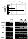
Fig. 1
Fig. 1. Paracrine signaling between myofibroblastic and ductular cells induced chemokine gene expression in ductular cells
(A) Chemokine related probe analysis Microarray analysis was performed in mono-/-co-culture samples derived from 3 independent experiments. Each probe having an expression ratio (average expression in co-cultures/average expression in monocultures) above 1.500 and below 0.666, was considered for the gene ontology (GO) analysis and assigned to its GO families. Probes belonging to Chemokine Activity, Chemokine receptor binding and Chemotaxis GeneOntology families are displayed as ratio of the expression in co-cultured vs mono-cultured cholangiocytes. (B) 2% Agarose gel of representative amplicon products QRT-PCR was performed in the murine cholangiocyte 603B line, rat NRC line and primary rat cholangiocytes using species-specific primers. Amplicon products were separated by electrophoresis on a 2% agarose gel buffered with 0.5× Tris-borate-EDTA (TBE) and then visualized using an AlphaImager 3400 Gel Analysis System. Ccl2/MCP-1 (chemokine C-C motif ligand 2/monocyte chemotactic protein-1); Ccl5 (chemokine C-C motif ligand 5); Ccl20/MIP3α (chemokine C-C motif ligand 20/macrophage inflammatory protein-3α), Cxcl1 (chemokine C-X-C motif ligand 1); Cxcl2 (chemokine C-X-C motif ligand 2); Cxcl5 (chemokine C-X-C motif ligand 5); Cxcl10 (chemokine C-X-C motif ligand 10); Cxcl11 (chemokine C-X-C motif ligand 11); Cxcl16 (chemokine C-X-C motif ligand 16).
Fig. 2
Fig. 2. Microarray data validation and comparison with freshly isolated cholangiocytes by QRT-PCR analysis
Chemokine gene mRNA levels were normalized to housekeeping gene expression in 603B mouse cholangiocytes or primary rat cholangiocytes. Data are displayed as fold-change in co-cultured 603B cholangiocytes relative to that in 603B mono-cultures (black bars), or in primary cholangiocytes isolated from 1 week BDL rat relative to sham controls (white bars). Data are representative of 3 experiments and shown as mean±SEM (*P<0.05, ** P<0.005).
Fig. 3
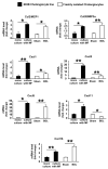
Fig. 3. Cholangiocyte chemokine gene induction is Hedgehog-dependent (A) QRT-PCR analysis of cholangiocytes cultured in myofibroblast-conditioned medium with or without Hh-neutralizing antibody
MF-HSC 8B were cultured for 6 days and conditioned medium was collected. This MF-HSC-conditioned medium was then added to monocultures of cholangiocyte cell line 603B that had been cultured without serum for 18 hours. Cholangiocytes were incubated with MF-HSC conditioned medium in presence of Hh-neutralizing antibody (5E1) (grey bars) or control IgG (black bars) (10 μg/mL) for an additional 24 hours. 603B cells cultured in unconditioned medium (white bars) provided an additional control group. QRT-PCR was then performed to analyze the expression of chemokine-related genes that had been altered when the cholagiocytes were co-cultured with MF-HSC line 8B. Data are representative of 3 independent experiments and expressed as mean±SEM. (*P<0.05, ** P<0.005) (B) QRT-PCR analysis of whole liver tissue from bile duct ligated (BDL) mice. Ptc-deficient (Ptc +/−) mice (black bars) (n=6) and their WT littermates (white bars) (n=6) underwent BDL and were analyzed for the same chemokines that were Hh-dependent in vitro. Data are shown as mean±SEM (*P<0.05, ** P<0.005).
Fig. 4
Fig. 4. Hh-dependent paracrine signaling caused ductular cells to secrete the NKT chemokine Cxcl16 and chemoattracted NKT cells in vitro
(A) Cxcl16 soluble protein analysis in primary cholangiocytes. Primary cholangiocytes were freshly isolated from sham or BDL rats. Supernatants were collected after 6 hours of incubation and analyzed for Cxcl16 by ELISA assay. (B) Effect of Hh ligand neutralization on Cxcl16 protein release by primary cholangiocytes. Freshly isolated cholangiocytes from normal rats were treated for 6 hours with either unconditioned medium (white bars) or 6 day MF-conditioned medium in presence of Hh ligand neutralizing antibody (5E1) (grey bars) or control IgG (black bars) (10μg/ml). Cxcl16 protein released into the supernatants was assessed by ELISA assay. (C) Cxcl16 soluble protein analysis in 603B cholangiocyte mono-/co-cultures. Conditioned medium was collected from 6 day mono- or co-cultures and Cxcl16 protein production was quantified by ELISA assay (D) Effect of Hh ligand neutralization on Cxcl16 protein release by 603B cholangiocytes. 6 day MF-conditioned medium was collected and added to mono-cultures of cholangiocytes in presence of Hh-neutralizing antibody (5E1) (grey bars) or control IgG (black bars) (10 μg/mL). 603B cells cultured with unconditioned medium (white bars) were used as controls. Supernatants were assessed by ELISA for Cxcl16 soluble protein levels, and cell pellets were analyzed by QRT-PCR for Adam10 gene expression. (E) (F) NKT cell migration assay. Cholangiocytes were cultured in absence/presence of MF-HSC. After 6 days, transwell filter inserts containing MF-HSC were removed. Cholangiocyte mono-/co-cultures were pre-treated for 1 hour with antibody neutralizing Cxcl16 or irrelevant IgG (5μg/mL). NKT cells were then added to the system using 5μm pore inserts, and their migration was monitored for 2 hours. Data are representative of 3 independent experiments and displayed as mean±SEM. (*P<0.05, ** P<0.005)
Fig. 5
Fig. 5. Cxcl16 localizes in Hh-responsive immature epithelial cells in PBC
(A–D) Double immunofluorescent staining. Cxcl16 (A) and CK-7 (B) co-localized (C–D) in representative sections from PBC liver. Normal liver co-staining failed to demonstrate CK-7/Cxcl16 double positive cells (C, NL insert). In all merged images, blue DAPI-staining demonstrates cell nuclei. (A–C) Original magnification ×40. (D) Magnified section of the indicated area in panel C. (E–F) QRT-PCR Analysis of whole liver tissue from13 patients with PBC and 7 control healthy livers (NL). (E) Cxcl16; (F) IFNγ. Data are expressed as as mean±SEM (*P<0.05 vs NL).

References

    1. Iredale JP. Models of liver fibrosis: exploring the dynamic nature of inflammation and repair in a solid organ. J Clin Invest. 2007;117:539–548. - PMC - PubMed
    1. Lazaridis KN, Strazzabosco M, Larusso NF. The cholangiopathies: disorders of biliary epithelia. Gastroenterology. 2004;127:1565–1577. - PubMed
    1. Marra F. Chemokines in liver inflammation and fibrosis. Front Biosci. 2002;7:d1899–1914. - PubMed
    1. Henderson NC, Iredale JP. Liver fibrosis: cellular mechanisms of progression and resolution. Clin Sci (Lond) 2007;112:265–280. - PubMed
    1. Omenetti A, Porrello A, Jung Y, Yang L, Popov Y, Choi SS, Witek RP, Alpini G, Ventr J, Vandongen HM, Syn W, Svegliati Baroni G, Benedetti A, Schuppan D, Diehl AM. Hedgehog signaling regulates epithelial-mesenchimal transition during biliary fibrosis in rodents and humans. JCI. 2008 - PMC - PubMed

Publication types

MeSH terms