. 2009 Sep;50(3):844-51.
doi: 10.1002/hep.23059.

Enhanced liver regeneration following changes induced by hepatocyte-specific genetic ablation of integrin-linked kinase

Affiliations

Enhanced liver regeneration following changes induced by hepatocyte-specific genetic ablation of integrin-linked kinase

Udayan Apte et al. Hepatology. 2009 Sep.

Abstract

Following liver regeneration after partial hepatectomy, liver grows back precisely to its original mass and does not exceed it. The mechanism regulating this "hepatostat" is not clear and no exceptions have been found to date. Although pathways initiating liver regeneration have been well studied, mechanisms involved in the termination of liver regeneration are unclear. Here, we report that integrin-linked kinase (ILK) (involved in transmission of the extracellular matrix [ECM] signaling by way of integrin receptors) and/or hepatic adaptations that ensue following ILK hepatocyte-targeted removal are critical for proper termination of liver regeneration. Following partial hepatectomy (PHx), mice with a liver-specific ILK ablation (ILK-KO-Liver) demonstrate a termination defect resulting in 58% larger liver than their original pre-PHx mass. This increase in post-PHx liver mass is due to sustained cell proliferation driven in part by increased signaling through hepatocyte growth factor (HGF), and the beta-catenin pathway and Hippo kinase pathways.

Conclusion: The data indicate that ECM-mediated signaling by way of ILK is essential in proper termination of liver regeneration. This is the first evidence of a defect leading to impaired termination of regeneration and excessive accumulation of liver weight following partial hepatectomy.


Figures

Figure 1
Figure 1
The data show the percentile of liver weight regained after partial hepatectomy in the control and ILK-KO-Liver mice. Following partial hepatectomy, the liver weights of Control and ILK-KO-Liver mice drop down to 54% and 53% of the original, respectively. At Day 14 after PHx, the liver weight of the control mice reaches 100% of the original whereas that of ILK-KO-Liver mice reaches 158% of the pre-hepatectomy weight. The data show the mean of at least 3 animals per point and are entirely derived from the data of Table 1. Statistical significance of differences is also shown in Table 1.
Fig. 2
Fig. 2
A. Percent of PCNA positive cells in the livers of ILK-KO-Liver and Control mice at various time points after PHx. ILK-KO-Liver mice have sustained excess cell proliferation at 5, 7 and 14 days post-PHx. The data show the mean and standard error from observations from three separate animals. The asterisks (*) indicate differences that are statistically significant at p<0.05. (B), Representative photomicrographs of PCNA immunohistochemistry in Control mice and ILK-KO-Liver mice at 14 days after PHx demonstrate prolonged proliferation of hepatocytes in livers of ILK-KO-Liver mice.
Figure 3
Figure 3
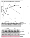
Changes in HGF and its receptor cMet and levels of total and nuclear beta catenin after partial hepatectomy. In A, data indicated expression values of HGF RNA in pooled samples of three animals per point. (CO: Control mice). B and C show representative western blots of HGF, Met and beta catenin (total (whole cell) and nuclear) from at least three experiments. There is increase in cMet at days 7 and 14 and increase in nuclear beta catenin at Days 2-7 post PHx in livers of ILK-KO-Liver mice compared to Control (CO).
Figure 4
Figure 4
Changes in cell cycle related proteins in ILK-KO-Liver mice after PHx, compared to Control. A and B: Western blots of total and phospho- AKT and total and phospho-p38 MAPK at different times after PHx. (C) Western blot analysis of c-myc and p-27 showing higher levels of c-myc and lower levels of p27 in ILK-KO-Liver mice. (D) Western blot showing no significant differences in TGFβ1 protein at 5, 7 and 14 days after PHx between ILK-KO-Liver and Control mice. (E) Western blot analysis of total YAP protein and phosphorylated YAP protein in ILK-KO-Liver and Control mice after PHx. At days 5, 7 and 14 after PHx there is an increase in total YAP and a decrease in the phospho-YAP to total YAP ratio in the ILK-KO-Liver mice. In D, representative photomicrographs of immunohistochemistry for YAP in Control and ILK-KO-Liver mice at 14 days post-PHx indicating higher levels of cytoplasmic and nuclear YAP protein in ILK-KO-Liver mice. Data expressed as western blots are representative of at least three separate experiments.
Figure 5
Figure 5
The top 120 expressed genes in time 0 (pre-PHx) in ILK-KO mice were identified by ranking of expression values in the Affymetrix gene array results. (for details, please supplement of reference (8)). The figure shows the expression of these genes at different times after PHx in both Control and ILK-KO-Liver mice. Gene expression values were temporarily increased fomr some of the genes, but they all returned closed to pre-hepatectomy values for Control mice. The expression values for ILK-KO-Liver mice however increased and for most of the genes remained elevated at Day 14 after PHx. Selenoprotein P had the most elevated expression in ILK-KO-Liver. MUP1 and Hemopexin (the highest expressions in Control at Day 7) also remained elevated at Day 14 in the ILK-KO-Liver group.

References

    1. Michalopoulos GK, DeFrances MC. Liver regeneration. Science. 1997;276:60–66. - PubMed
    1. Michalopoulos GK. Liver regeneration. J Cell Physiol. 2007 - PMC - PubMed
    1. Fausto N, Campbell JS, Riehle KJ. Liver regeneration. Hepatology. 2006;43:S45–53. - PubMed
    1. Mohammed FF, Pennington CJ, Kassiri Z, Rubin JS, Soloway PD, Ruther U, Edwards DR, et al. Metalloproteinase inhibitor TIMP-1 affects hepatocyte cell cycle via HGF activation in murine liver regeneration. Hepatology. 2005;41:857–867. - PubMed
    1. Oe S, Lemmer ER, Conner EA, Factor VM, Leveen P, Larsson J, Karlsson S, et al. Intact signaling by transforming growth factor beta is not required for termination of liver regeneration in mice. Hepatology. 2004;40:1098–1105. - PubMed

MeSH terms