Review
. 2010:26:363-96.
doi: 10.1146/annurev.cellbio.042308.113238.

Rolling cell adhesion

Affiliations
Review

Rolling cell adhesion

Rodger P McEver et al. Annu Rev Cell Dev Biol. 2010.

Abstract

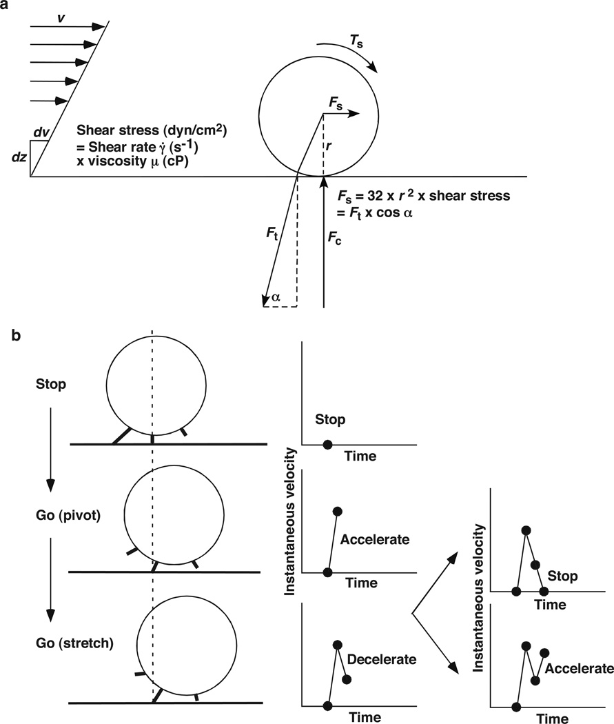
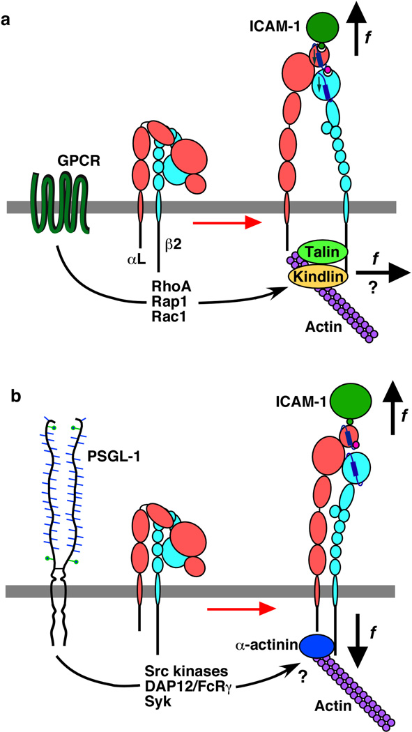
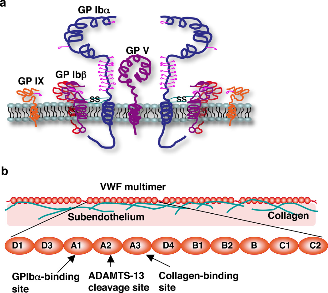
Rolling adhesion on vascular surfaces is the first step in recruiting circulating leukocytes, hematopoietic progenitors, or platelets to specific organs or to sites of infection or injury. Rolling requires the rapid yet balanced formation and dissociation of adhesive bonds in the challenging environment of blood flow. This review explores how structurally distinct adhesion receptors interact through mechanically regulated kinetics with their ligands to meet these challenges. Remarkably, increasing force applied to adhesive bonds first prolongs their lifetimes (catch bonds) and then shortens their lifetimes (slip bonds). Catch bonds mediate the counterintuitive phenomenon of flow-enhanced rolling adhesion. Force-regulated disruptions of receptor interdomain or intradomain interactions remote from the ligand-binding surface generate catch bonds. Adhesion receptor dimerization, clustering in membrane domains, and interactions with the cytoskeleton modulate the forces applied to bonds. Both inside-out and outside-in cell signals regulate these processes.


Figures

Figure 1
Figure 1
Parameters of tethering under flow. (a) The fluid velocity v of a Couette flow field bordered with a solid surface (x-y plane) is parallel to the surface and increases linearly with the distance away from the surface (z direction). The shear rate γ̇ = dv/dz is reciprocal to the slope of the velocity profile. Fluid mechanics theory predicts that the translational velocity V and angular velocity Ω of a sphere (or cell) of radius r freely moving above a surface in an otherwise Couette flow are proportional to rγ̇ and r, respectively (Goldman et al 1967). The sphere bottom has a sliding velocity VsVrΩ ∝ r relative to the surface (Chang & Hammer 1999). Adapted with permission from (Yago et al 2007). (b) Faster sliding velocity increases the number of surface ligands that an adhesion receptor on the flowing cell contacts per unit time. (c) Faster sliding velocity reduces the time that the receptor contacts the ligand before it moves away.
Figure 2
Figure 2
Parameters of rolling adhesion under flow. (a) The rolling motions of a sphere or cell of radius r are governed by the balance of the resultant force Fs and torque Ts exerted by the flowing fluid, the tether force Ft applied through the receptor-ligand bond, and the contact force Fc. The conversion of wall shear stress to Ft using the indicated variables is described in (Yago et al 2004). (b) A rolling sphere stops when the adhesive bond sustains the full load required to balance the maximum Ft and Ts. After the bond dissociates, the sphere accelerates as it pivots on a newly formed bond downstream and then decelerates as force develops in the bond. The sphere stops again if the new bond has sufficient strength to withstand the full load and lives long enough to survive loading, or it accelerates if the bond dissociates prematurely. Both panels adapted with permission from (Yago et al 2004).
Figure 3
Figure 3
Multistep leukocyte adhesion cascade. Selectins initiate tethering and rolling of leukocytes. Depending on their activation state, integrins mediate slower rolling or cause the cells to arrest. Integrins also mediate spreading, crawling, and migration between or through endothelial cells into the underlying tissues.
Figure 4
Figure 4
Selectins and their major glycoprotein ligands. The upper inset depicts the N-terminal glycosulfopeptide region of human PSGL-1 that binds to P-selectin (and L-selectin). The lower insert depicts an example of a sialylated, fucosylated, and sulfated O-glycan on PNAd mucins that binds to L-selectin.
Figure 5
Figure 5
Force-dependent lifetimes of single bonds between PSGL-1 and P-selectin or L-selectin. Lifetimes of transient neutrophil tethers to low-density P-selectin or L-selectin at different wall shear stresses were measured by video microscopy. Adapted with permission from (Marshall et al 2003, Sarangapani et al 2004).
Figure 6
Figure 6
Bent and extended selectin structures. (a) Overlay of the lectin and EGF domains of P-selectin in its bent and extended forms. The magenta and blue spheres at the top represent the respective Ca2+ ion on each structure. (b) Binding of fucose (part of the sLex binding determinant) to the bent and extended forms of P-selectin. The dashed black lines represent interactions of the Ca2+ ion with residues on P-selectin. The dashed red lines represent interactions of the fucose with the Ca2+ ion or residues on P-selectin. (c) Relative orientations of Y37 in the lectin domain and G138 in the EGF domain in the bent and extended forms of P-selectin. (d) Relative orientations of Y37 in the lectin domain and N138 in the EGF domain in the bent and extended forms of L-selectin. The black dashed line indicates a hydrogen bond. [From Protein Data Bank (PDB) ID codes 1G1Q, 1G1R, 1G1S, and 3CFW (Klopocki et al 2008, Somers et al 2000).] The extended structure of L-selectin was derived by molecular modeling (Lou et al 2006).
Figure 7
Figure 7
Models for catch bonds. (a) Allosteric model. (b) Sliding-rebinding model.
Figure 8
Figure 8
Flow-enhanced rolling adhesion. Neutrophils were perfused over immobilized PSGL-1 at the indicated wall shear stress. The tethering rate (a), mean rolling velocity (b), and number of cells rolling per field (c) were measured. Adapted with permission from (Yago et al 2004, Yago et al 2007).
Figure 9
Figure 9
Schematic showing tensile forces (Ft) between cells in a flowing doublet as well as between a cell and the wall under a simple shear field. <Ft> indicates average force over a cycle of tumbling.
Figure 10
Figure 10
Effects of cell-surface organization on selectin-ligand interactions under flow. (a) Extension of a long membrane tether from a microvillus after disruption of cytoskeletal connections with the membrane. (b) P-selectin and PSGL-1 form dimers. P-selectin clusters in clathrin-coated pits through interactions of its cytoplasmic domain. PSGL-1 associates with lipid rafts and clusters in microvilli, perhaps indirectly through interactions of other raft components with the cytoskeleton. Note that the tip of a microvillus is actually larger than a clathrin-coated pit, and PSGL-1 molecules in different regions of the tip may interact with P-selectin molecules in two or more clustered pits. (c) L-selectin clusters in microvilli through direct interactions of its cytoplasmic domain with α-actinin and ERM proteins, which connect to actin filaments.
Figure 11
Figure 11
Domain organization of integrins and ligands that mediate leukocyte rolling or arrest. (a, b) Rearrangement of domains during activation of integrin α4β1, which lacks an α I domain (a) or during activation of integrin αLβ2, which has an α I domain (b). The domains of each headpiece are indicated. Binding of the N-terminal Ig domain of VCAM-1 (a) or ICAM-1 (b) to the respective headpiece is shown; for clarity, the other Ig domains of VCAM-1 or ICAM-1 are shown only in panel d. (c) Binding of the small-molecule antagonist XVA143 to the β I domain of αLβ2.
Figure 12
Figure 12
Overlay of the structures of low-affinity, intermediate-affinity, and high-affinity conformations of the αL I domain. Regions of minimal conformational changes in all three conformations are shown in gray. The introduced disulfide bonds that stabilize each conformation and the movements of the MIDAS metal ion (sphere), the α1-loop, and the α7-helix are color coded as shown. The directions of movement are depicted with arrows. [From PDB ID codes 1ZOO, 1MJN, and 1TOP (Qu & Leahy 1996, Shimaoka et al 2003, Song et al 2005).]
Figure 13
Figure 13
Differential activation of integrin αLβ2 to mediate leukocyte arrest or rolling. (a) Chemokine binding to its G protein-coupled receptor (GPCR) activates Rho-family GTPases and effectors that recruit talin and kindlins to the β2 tail, which in turn bind to actin filaments. Lateral forces exerted by the cytoskeleton may separate the integrin legs, favoring integrin extension, swing-out of the hybrid domain, and allosteric conversion of the αL I domain to its high-affinity conformation. Tensile force applied to ICAM-1 bound to the αL I domain may further stabilize this conformation, causing leukocyte arrest. (b) Selectin engagement of PSGL-1 on rolling leukocytes propagates signals upstream and downstream of Syk. These signals might permit binding of α-actinin to the β2 tail. Incomplete leg separation may cause the integrin to extend in a conformation that resists force-induced conversion of the αL I domain to its high-affinity state.
Figure 14
Figure 14
Rolling of platelets on vascular surfaces. (a) After vascular injury, platelets tether to and roll (translocate) on subendothelial matrices through interactions of platelet GPIbα with VWF. Signaling during rolling activates platelet β1 and β3 integrins, which mediate arrest and aggregation. (b) Unstimulated platelets roll on P-selectin on activated endothelial cells, probably through interactions with GPIbα. Activated platelets use P-selectin to roll on PNAd or PSGL-1 expressed on some endothelial cells and on PSGL-1 expressed on leukocyte fragments bound to the endothelial cell surface. Activated platelets bound to leukocytes roll together on endothelial cells.
Figure 15
Figure 15
Schematic of the platelet GPIb/IX/V complex (a) and VWF (b).
Figure 16
Figure 16
Structural model for catch bonds between GPIbα and VWF. (a–e) Sequential snapshots of steered MD-simulated structures showing pathways of the sliding-rebinding mechanism. Mauve, A1; gray, GPIbαN; red spheres, D1269; blue spheres, R1306; green spheres, R1450; blue β-strands, β-switch of GPIbα; red β-strands, central β-sheet of A1; red sticks, E14; blue sticks, R1334. The equilibrated structure (a) was used as a starting point of steered MD simulations to generate the simulated structures in b–e. (f) Overlay of the structures in a (colors same as in a) and d (green, A1; cyan, GPIbα), showing the rotation of A1 and sliding of the GPIbα β-finger over A1. (g) Sliding-rebinding mechanism for GPIbα/VWF A1 catch bonds. Adapted with permission from (Yago et al 2008).

References

    1. Abitorabi MA, Pachynski RK, Ferrando RE, Tidswell M, Erle DJ. Presentation of integrins on leukocyte microvilli: a role for the extracellular domain in determining membrane localization. J Cell Biol. 1997;139:563–571. - PMC - PubMed
    1. Alon R, Chen SQ, Puri KD, Finger EB, Springer TA. The kinetics of L-selectin tethers and the mechanics of selectin-mediated rolling. J Cell Biol. 1997;138:1169–1180. - PMC - PubMed
    1. Alon R, Feigelson SW, Manevich E, Rose DM, Schmitz J, et al. alpha4-beta1-dependent adhesion strengthening under mechanical strain is regulated by paxillin association with the alpha4-cytoplasmic domain. J Cell Biol. 2005;171:1073–1084. - PMC - PubMed
    1. Alon R, Hammer DA, Springer TA. Lifetime of the P-selectin: carbohydrate bond and its response to tensile force in hydrodynamic flow. Nature. 1995a;374:539–542. - PubMed
    1. Alon R, Kassner PD, Carr MW, Finger EB, Hemler ME, Springer TA. The integrin VLA-4 supports tethering and rolling in flow on VCAM-1. J Cell Biol. 1995b;128:1243–1253. - PMC - PubMed

Publication types

LinkOut - more resources