. 2009 Jul 4:2:22.
doi: 10.1186/1757-1146-2-22.

A protocol for classifying normal- and flat-arched foot posture for research studies using clinical and radiographic measurements

Affiliations

A protocol for classifying normal- and flat-arched foot posture for research studies using clinical and radiographic measurements

George S Murley et al. J Foot Ankle Res. .

Abstract

Background: There are several clinical and radiological methods available to classify foot posture in research, however there is no clear strategy for selecting the most appropriate measurements. Therefore, the aim of this study was to develop a foot screening protocol to distinguish between participants with normal- and flat-arched feet who would then subsequently be recruited into a series of laboratory-based gait studies.

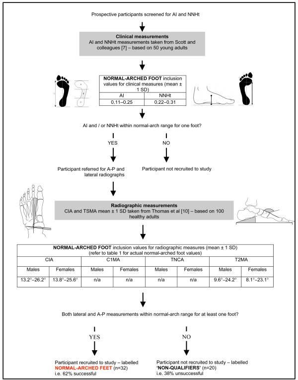
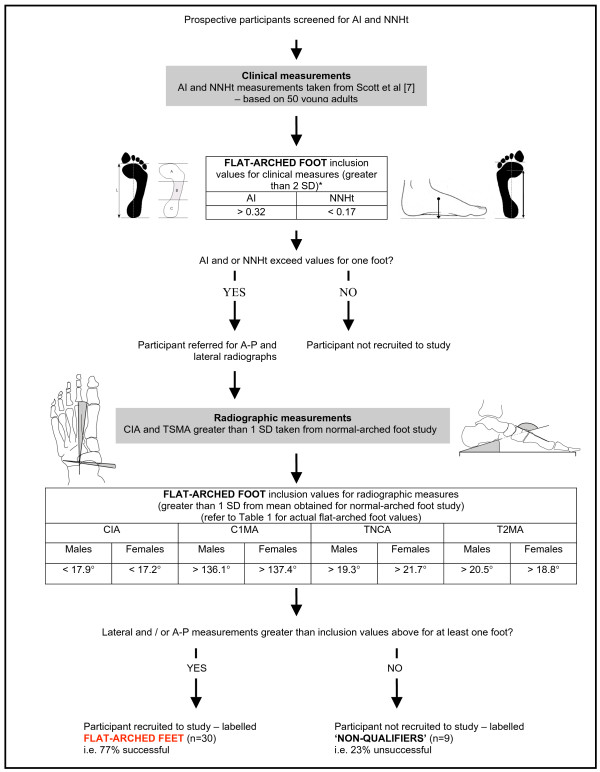
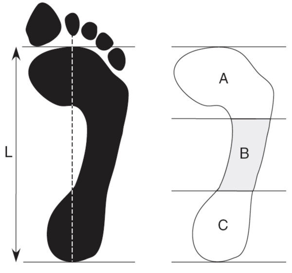
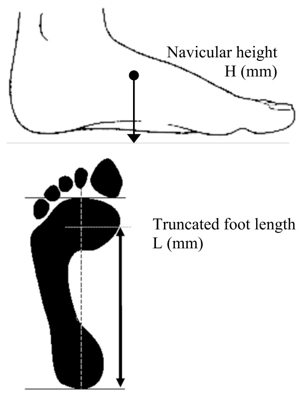
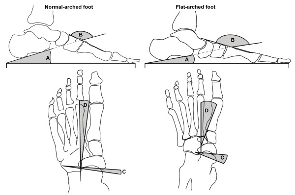
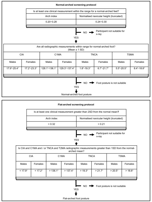
Methods: The foot posture of ninety-one asymptomatic young adults was assessed using two clinical measurements (normalised navicular height and arch index) and four radiological measurements taken from antero-posterior and lateral x-rays (talus-second metatarsal angle, talo-navicular coverage angle, calcaneal inclination angle and calcaneal-first metatarsal angle). Normative foot posture values were taken from the literature and used to recruit participants with normal-arched feet. Data from these participants were subsequently used to define the boundary between normal- and flat-arched feet. This information was then used to recruit participants with flat-arched feet. The relationship between the clinical and radiographic measures of foot posture was also explored.

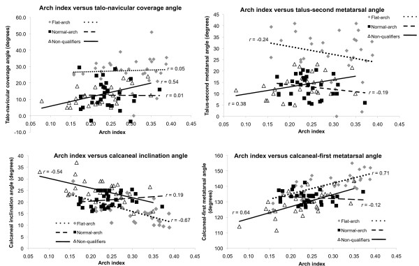
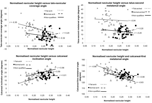
Results: Thirty-two participants were recruited to the normal-arched study, 31 qualified for the flat-arched study and 28 participants were classified as having neither normal- or flat-arched feet and were not suitable for either study. The values obtained from the two clinical and four radiological measurements established two clearly defined foot posture groups. Correlations among clinical and radiological measures were significant (p < 0.05) and ranged from r = 0.24 to 0.70. Interestingly, the clinical measures were more strongly associated with the radiographic angles obtained from the lateral view.

Conclusion: This foot screening protocol provides a coherent strategy for researchers planning to recruit participants with normal- and flat-arched feet. However, further research is required to determine whether foot posture variations in the sagittal, transverse or both planes provide the best descriptor of the flat foot.


Figures

Figure 1
Figure 1
Screening protocol for normal-arched foot posture. Flow chart shows how the foot posture screening protocol was derived from normative data. * Values derived from Scott and colleagues [7]. CIA – calcaneal inclination angle, C1MA – calcaneal-first metatarsal angle, TNCA – talo-navicular coverage angle, T2MA – talus-second metatarsal angle.
Figure 2
Figure 2
Screening protocol for flat-arched foot posture. Flow chart shows how the foot posture screening protocol was derived from normative data. * Values derived from Scott and colleagues [7]. The rationale for using 2 SD standard deviations was to increase the likelihood of participants with flat-arched feet qualifying for inclusion via radiographic appraisal. CIA – calcaneal inclination angle, C1MA – calcaneal-first metatarsal angle, TNCA – talo-navicular coverage angle, T2MA – talus-second metatarsal angle.
Figure 3
Figure 3
Arch index. Footprint with reference lines for calculating the arch index. The length of the foot (excluding the toes) is divided into equal thirds to give three regions: A – forefoot; B – midfoot; and C – heel. The arch index is then calculated by dividing the midfoot region (B) by the entire footprint area (i.e. Arch index = B/[A+B+C]).
Figure 4
Figure 4
Normalised navicular height (truncated). Calculating normalised navicular height truncated. The distance between the supporting surface and the navicular tuberosity is measured. Foot length is truncated by measuring the perpendicular distance from the 1st metatarsophalangeal joint to the most posterior aspect of the heel. Normalised navicular height truncated is calculated by dividing the height of the navicular tuberosity from the ground (H) by the truncated foot length (L) (i.e. Normalised navicular height truncated = H/L).
Figure 5
Figure 5
Radiographic measurements. Traces from two representative participants illustrate x-ray angular measurements from normal (left) and flat-arched (right) foot posture. Lateral views (top) show: calcaneal inclination angle; calcaneal-first metatarsal angle; anterior posterior views (bottom) show: talonavicular coverage angle; talus second metatarsal angle. A – calcaneal inclination angle, B – calcaneal-first metatarsal angle, C – talo-navicular coverage angle, D – talus-second metatarsal angle. Angle A decreases with flat-arched foot posture; angle B, C and D increase with flat-arched foot posture, compared to the normal-arched foot posture.
Figure 6
Figure 6
Arch index versus radiographic measures for each foot posture group. Scatter plots with trend lines for the arch index and radiographic measures of foot posture show the distribution of values for normal-arch, flat-arch and non-qualifying foot postures.
Figure 7
Figure 7
Normalised navicular height versus radiographic measures for each foot posture group. Scatter plots with trend lines for the normalised navicular height and radiographic measures of foot posture show the distribution of values for normal-arch, flat-arch and non-qualifying foot postures.
Figure 8
Figure 8
Screening protocol for normal- and flat-arched foot posture. Flow chart shows how the foot posture screening protocol can be applied to future studies recruiting participants with normal- and flat-arched foot posture. CIA – calcaneal inclination angle, C1MA – calcaneal-first metatarsal angle, TNCA – talo-navicular coverage angle, T2MA – talus-second metatarsal angle.

References

    1. Redmond AC, Crane YZ, Menz HB. Normative values for the Foot Posture Index. J Foot Ankle Res. 2008;2:6. doi: 10.1186/1757-1146-1-6. - DOI - PMC - PubMed
    1. Redmond AC, Crosbie J, Ouvrier RA. Development and validation of a novel rating system for scoring standing foot posture: The Foot Posture Index. Clin Biomech (Bristol, Avon). . 2006;2(1):89–98. doi: 10.1016/j.clinbiomech.2005.08.002. - DOI - PubMed
    1. Hunt AE, Smith RM. Mechanics and control of the flat versus normal foot during the stance phase of walking. Biomech (Bristol, Avon) 2004;2(4):391–397. doi: 10.1016/j.clinbiomech.2003.12.010. - DOI - PubMed
    1. Murley GS, Landorf KB, Menz HB, Bird AR. Effect of foot posture, foot orthoses and footwear on lower limb muscle activity during walking and running: a systematic review. Gait Posture. 2009;2:172–187. doi: 10.1016/j.gaitpost.2008.08.015. - DOI - PubMed
    1. Brewerton DA, Sandifer PH, Sweetnam DR. "Idiopathic" Pes Cavus: an Investigation into Its Aetiology. Br Med J. 1963;2:659–661. doi: 10.1136/bmj.2.5358.659. - DOI - PMC - PubMed